Michael Zucker MD
- Professor of Clinical Radiology, Emeritus, David Geffen School of Medicine at
- University of California Los Angeles, Los Angeles, California
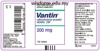
Vantin dosages: 200 mg, 100 mg
Vantin packs: 30 pills, 60 pills, 90 pills, 120 pills, 180 pills, 270 pills
Order vantin with paypal
It is neces- sary to use the traction countertraction concept xifaxan antibiotic ibs purchase genuine vantin on-line, whereby the surgeon pulls on one side and the assistant asserts gentle traction on the opposite side to put the tissue under tension antibiotic resistance guidelines buy vantin mastercard. The patient is placed in the supine position in such a way that the team can move around Right the patient with ease virus black muslim in the white house vantin 100 mg order online. The surgeon stands on the patient’s left, watching the monitor on Hemicolectomy the other side (Fig. The operation can be performed in two fashions: medial to lateral or lateral to medial. In the medial to lateral technique, the peritoneal adhesions of the colon to the abdominal wall are used as counter-traction while dissecting and dividing the ileocolic vessels. The patient is placed in Trendelenberg and right side up to remove the small bowel from pelvis and right lower quadrant. First, the terminal ileum is grasped with the left hand and pulled towards the anterior abdominal wall placing tension on the ileo- colic vessels. For this, the patient is put in the Trendelenburg, right side up position, putting the cecum under tension and facilitating the dissection. Once again, this puts the appropriate tension on the hepatic fexure to assist the dissection. The mesocolon should now be clearly identifable, and if the patient is not too obese, it is possible to perform intra-abdominal division of the vessels with vascular staplers. Otherwise, if mobilization of the colon is suffcient, it is possible to deliver the whole right colon and the terminal ileum through a right upper quadrant muscle splitting incision, followed by an anasto- mosis outside the abdomen. If a hand assisted port is used, a midline incision is used for the hand port, which can be used to deliver the colon and construct the anastomosis at the end of the case. A umbilical scope; B surgeon’s left hand; C surgeon’s right hand; D, E graspers of the frst assistant. Note that the trocar Left positions are moved down when a low anterior resection is performed. Hemicolectomy As described previously, a medial to lateral or lateral to medial approach can be cho- sen. In the lateral to medial approach, the frst step is to mobilize the sigmoid colon by applying traction and counter-traction during the dissection. At this point it is important to identify the rectosigmoid junction and the ureters. If a ureter is not clearly visible because of intense infammation, it is possible to locate it by inserting a ureteral stent or even an ultraviolet stent. One trick is to move the camera to one of the left lower ports in order to get a direct view of the left fascia of Toldt. Again, traction on the mesocolon of the transverse colon and traction on the adhesions of the splenic fexure will lead to safe division of the splenic fexure. The spleen should not be seen and one should stay as close as possible to the colon (Fig. When the whole colon has been mobilized, it is possible to go down into the pelvis and decide on the site for the anastomosis. Metallic clips are avoided as they may interfere with proper fring of the stapler. Sometimes, with a large rectum it is necessary to fre two shots to complete the transection. Before fring the stapler it is essential to make sure that no rectal tubes are in the rectum. The fully mobilized left colon is exteriorized through a muscle splitting incision using one of the left lower quadrant ports or a Pfannenstiel, and the specimen is resected. An anvil is then placed in the proximal end after trimming the area appropriately, and a purse string suture applied. A circular stapler is then introduced into the rectum, with care being taken to perforate anterior to the staple line. Using a specifc instrument that allows appropriate handling of the anvil, it is connected to the shaft of the stapler and fred (Fig.
200 mg vantin purchase otc
The two methods most commonly used to identify a pulmonary vein source of spontaneous impulse formations are use of the electrocardiogram and use of mapping catheters along the posterior septum or anterolateral right atrium with the proximal poles along the posterior-superior septum antimicrobial bar soap discount vantin online american express, and within the coronary sinus antibiotic side effects 200 mg vantin purchase. The proximity to the posterior superior septum infection from pedicure order vantin 100 mg fast delivery, with early breakthrough to the right atrium, and simultaneous activation of the left atrium leads to this P-wave morphology. In contrast, atrial impulses arising from left superior pulmonary vein foci have broader P waves because they are far removed from the septum. They are frequently broad and notched and demonstrate total positivity in lead V as1 well as being positive in the inferior leads (lead 3 usually being more positive than lead 2). In contrast to activation associated with foci originating in the right superior pulmonary vein, the left superior vein foci produces activation of the coronary sinus earlier than the right atrial catheter, particularly when the coronary sinus catheter is positioned with its distal pole anteriorly. These two observations can provide evidence for a pulmonary vein origin for the spontaneous ectopy and initiation of atrial fibrillation, and justify P. Of note, negative P waves in lead 1 are never seen in left or right pulmonary vein foci, since the pulmonary veins are posterior in the chest. A negative P wave in lead 1 suggests a left atrial appendage or lateral mitral annulus origin. As such, I recommend that no sedative be given when one is attempting to demonstrate spontaneous atrial impulse formation. Moreover, it is most commonly necessary to add pharmacologic agents to facilitate impulse formation. Because of the unreliability of seeing all potential sources of pulmonary vein foci, we do not rely on them for ablation of atrial fibrillation. An ectopic atrial arrhythmia was initiated from that ablation/mapping catheter as demonstrated by earliest activation from the distal tip of the ablation catheter with subsequent activation to the proximal portion of the pulmonary vein associated with a layer left atrial component. Thus, initiation of atrial tachyarrhythmias from the pulmonary veins by mechanical induction of catheter introduction itself can produce misleading rhythms that should not be targeted for ablation. Once the appearance of pulmonary vein foci has been suggested, transseptal catheterization should be performed. Although some investigators use a single transseptal puncture followed by introduction of two guidewires and then reintroduction of two sheaths, I prefer two (or occasionally three) separate transseptal procedures. I believe the latter approach is associated with less femoral vein bleeding and local complications as well as a lower incidence of persistent atrial septal defect following a procedure. Intracardiac ultrasound or transesophageal echocardiography may also be employed to facilitate catheter positioning. Once the left atrium is entered the goals for the procedure are to identify vein and/or branch of the vein from which the impulse arises and localize either the earliest site of activation within that vein or branch, or the segment from which the earliest activation is recorded. The initial method used was point-by-point activation mapping in the pulmonary veins. This method is still useful, particularly using electroanatomic mapping, which allows one to tag the anatomic location of individual potentials. The initiation of atrial ectopic impulse formation in the pulmonary veins only after a catheter has been introduced into the pulmonary veins does not signify that the source of atrial fibrillation has been identified. The catheter should be withdrawn to assess whether or not this impulse formation exists spontaneously. This is in contrast to an ongoing atrial tachyarrhythmia in which a pulmonary vein is explored and early activation found (Fig. One of the other clues that this rhythm was not flutter is simultaneous activation from high to low on the lateral right atrial free wall and on the septum. Ablation of the focal tachycardia, which was initially performed in the pulmonary vein, is now done by targeting the exit site from the vein at the ostial cuff, or more commonly, by isolating the entire vein. The halo catheter distally records the lateral isthmus and proximally the superior septum. The isthmus catheter is adjacent to the halo catheter distal, which was at the site of a prior ablation for proven isthmus-dependent flutter. The split electrograms are at the site of block produced by the prior linear lesion. The activation in the left superior pulmonary vein records a pulmonary vein potential of 130 msec prior to the P wave with a later left atrial potential (A*). Subsequent ablation of the pulmonary vein focus eliminated the need for antiarrhythmic therapy and the patient has been free of arrhythmia since.
Syndromes
- Foul-smelling vaginal discharge
- Redness or swelling of the ankle joint
- Treatment is started in the fall or early winter, before the symptoms of SAD begin.
- Difficulty breathing
- Transderm-Nitro
- Wheezing
- If the body is pink and the extremities are blue, the infant scores 1 for color.
Vantin 200 mg buy fast delivery
The skin has deep rugae where the skin is rubbing on underwear secondary to vaginal prolapse antibiotics xorimax cheap vantin online. It suggests that women should be assessed with an empty bladder and ideally an empty rectum because an increased bladder volume can restrict the degree of descent of the vaginal prolapse virus del papiloma humano 100 mg vantin purchase mastercard. Any cystocele antimicrobial yarn suppliers order vantin overnight, rectocele, uterine, or vault descent can be best assessed in the Sims’ (left lateral) position on coughing and straining using a Sims’ speculum. Alternatively, examination may also be made in lithotomy using the lower blade of a Graves’ speculum to assist in assessing the opposite vaginal wall [31]. Women may also be examined standing and this may be the only way for a vaginal prolapse to be demonstrated successfully [32]. After the examination for pelvic organ prolapse, the clinician should report the degree of prolapse in terms of the descent of the anterior and posterior vaginal walls, as well as the cervix, or in the case of a woman who has had a hysterectomy, the vaginal vault. Various staging or grading methods have been described and the level of the hymen is usually used. The pelvic organ prolapse quantification system is a validated method that involves using a measuring device marked in centimeters to measure specific points on the anterior and posterior vaginal wall as well as the descent of the cervix or vaginal cuff allowing quantification of the degree of vaginal prolapse [33]. Women can be reproducibly examined in the left lateral and standing positions [32]. The vagina should also be examined for evidence of atrophy, inflammation, pain, or scarring from previous surgical procedures. Other aspects of pelvic examination include examination of the urethra where urethral mucosal prolapse or a caruncle may be seen. A urethral diverticulum may present with a midline anterior vaginal wall swelling or with tenderness over the length of the urethra as well as postmicturition leakage. If a woman is complaining of a discharge or has had a recent onset of symptoms of urgency and frequency, it may be useful to obtain swabs to culture for Chlamydia, atypical organisms, or gonococcus. A bimanual examination should be performed to exclude abnormal pelvic organs, masses, or uterine impaction and can exclude a large postmicturition urinary residual. Pelvic masses such as ovarian cysts and uterine enlargement greater than 12 weeks size can cause pressure symptoms on the bladder and rectum resulting in urinary frequency or retention; often, the symptoms resolve once the mass has been removed. If pelvic pain is a problem, it is important that a digital examination is performed to assess where the pain originates and in particular to determine trigger points in the levator ani and also tenderness upon palpation of the pelvic organs adjacent to the vagina. Finally, rectal examination is particularly important in the elderly to exclude fecal impaction, which can aggravate urinary incontinence. The method in which symptoms are ascertained may alter the results and self-completed questionnaires seem to be the best. It is important to use reproducible and validated questionnaires as symptoms alone have become the basis of conservative management and have been suggested as indicators for the surgical treatment of pure stress incontinence. In some cases, an obvious cause can be found and treated, thus potentially avoiding the need for further investigations. Diuresis pattern, plasma vasopressin and blood pressure in healthy elderly persons with nocturia and nocturnal polyuria. The relationship between urinary symptom questionnaires and urodynamic diagnoses: An analysis of two methods of questionnaire administration. Multinational study of reliability and validity of the King’s Health Questionnaire in patients with overactive bladder. Definition and classification of urinary incontinence: Recommendations of the urodynamic society. Urinary incontinence in French women: Prevalence, risk factors, and impact on quality of life. The tension free vaginal tape operation for women with mixed incontinence: Do preoperative variables predict the outcome? Urinary incontinence at orgasm: Relation to detrusor overactivity and treatment efficacy. Incontinence and detrusor dysfunction associated with pelvic organ prolapse: Clinical value of preoperative urodynamic evaluation. Role of alpha2-adrenoceptors and glutamate mechanisms in the external urethral sphincter continence reflex in rats.
Cheap 100 mg vantin
This suggests that differences in autonomic tone make this measurement inherently variable klebsiella antibiotic resistance mechanism order generic vantin from india. No data are available in an age-matched infection zombie movies purchase generic vantin on line, control population analyzing sinus node electrograms after autonomic blockade antibiotics for acne list order vantin from india. The onset of carotid sinus massage as well as the presence of a blocked sinus node depolarization (labeled S-A exit block) and A-V nodal block of the following atrial depolarization (right-hand end of tracing) during carotid sinus massage. Note also the presence of the atrial repolarization waves (upward heavy arrows) and minimal manifestation of ventricular T waves (heavy downward arrows). T-wave activity is absent following the blocked atrial depolarization (right end of tracing). This is most likely the result of prolonged perinodal refractoriness, which prohibits premature impulses from penetrating and resetting the sinus node. Another response more commonly noted in patients with clinical sinus dysfunction is the progressive lengthening 66 67 of A1-A3, resulting in either abbreviation or abolition of the plateau portion of the curve. The increase probably results from improvement in retrograde conduction into the sinoatrial node, resulting in depression of automaticity. Sinus Node Recovery Time 68 Suppression of pacemaker activity by driving the heart at a faster rate was first noted by Gaskell. The mechanism of overdrive suppression of the sinus node remains unclear; factors P. They include (a) proximity of the stimulation site to the sinus node, (b) local concentrations of acetylcholine and norepinephrine, and (c) conduction time into and out of the sinus node. It is imperative that at least 1 minute be allowed between paced cycle lengths to ensure full recovery of the sinus node. High- and low-atrial electrograms are usually simultaneously recorded to enable one to determine whether escape beats at the termination of pacing originate from the sinus node. Confirmation of the sinus node as the origin of the escape beats depends on demonstration of a similar P-wave morphology and atrial activation sequence to that observed during sinus rhythm before atrial pacing. Changes in P-wave morphology and/or atrial activation sequence suggest a shift of pacemaker. Such shifts represent a limitation to all indirect methods of assessing sinus node function. In addition, the presence of junctional escapes and sudden unexpected pauses during the recovery period should be noted. The marked differences in reported “normal” values probably reflect differences in patient populations with respect to autonomic tone and structural cardiac disease as well as to differences in methodology. This prolongation may occur when the spontaneous sinus cycle length is less than 800 msec and thus should not be used in such circumstances. Thus, when sinus node recovery from overdrive suppression is evaluated, some consideration must be given to the basic sinus cycle length. Normally, there is gradual warm-up (shortening of cycle length) following the cessation of overdrive pacing until the control sinus length is achieved (see Fig. It is not unusual to find oscillation of recovery cycle lengths before full recovery, but the pattern of oscillation of sinus cycle length shortening should fall within the limits described 77 10 42 54 74 76 77 by Benditt et al. This sophisticated technique is, however, limited by (a) effects of change in autonomic tone that are due to the hemodynamic consequences of pacing; (b) changes in P-wave morphology suggesting a change in pacemaker location and/or exit; (c) sinoatrial entrance block; and (d) secondary pauses. These are also limitations common to all methods analyzing the response to overdrive suppression. Sudden and marked secondary pauses occurring during sinus recovery are abnormal and in most instances reflect changes in sinoatrial conduction; however, a change in automaticity with or without pacemaker shift is also possible. Because these secondary pauses represent abnormalities of sinus function and because they occur more frequently following rapid pacing, atrial pacing should be carried out at rates up to 200 bpm. Several investigators have demonstrated sinoatrial exit block of variable duration to be the primary mechanism of prolonged pauses with a lesser component of depression of automaticity (Fig. This can clearly be demonstrated by persistence of sinus node electrograms during pauses. It is impossible to predict to what extent sinoatrial conduction and depression of automaticity contribute to the pauses noted in any individual, and only through the use of sinus node electrograms can this be adequately assessed. Following the last stimulated complex of overdrive pacing (s, arrow), a long pause ensues.
Vantin 100 mg order without prescription
Septal cartilage is pre- can reduce convexity is emphasized in this example where it is applied ferred 606 antibiotic vantin 200 mg overnight delivery. However antibiotic resistance food chain generic 100 mg vantin mastercard, it is usually unavailable in sufficient over the superficial surface of collapsed upper lateral cartilages quantities infection near fingernail vantin 200 mg purchase on line. Since it can be straightened and stiffened as described above, it is useful for almost every situation where septal cartilage would ordinarily be used. However, when large amounts of 7 6 5 4 3 2 Increases by Decreases 1 35% by 48% Fig. It does not preclude the No points With points Cartilage incision need for spreader grafts and flaps, however, which are the main means of maintaining internal valve integrity F i g. Scoring the cartilage to achieve the same degree of straightness can reduce the cartilage strength by 50 % Fig. Erol [6] demonstrated that diced cartilage in a ary cases is the use of irradiated allograft rib cartilage [20]. However, we have This can be useful when the patient is resistant to harvesting remained with fascia to avoid the possibility of absorption autogenous material. Although some reports into small bits approximately 1 mm in size and placed in a of long-term survival exist [3–10], one must be prepared for blanket of fascia (on a silicone block). An option we never consider for secondary rhinoplasty is the use of implants of any material. Complications from implants are much more difficult to correct and therefore are not used. Prior to harvesting ear cartilage, an ellipse of the skin is deepithelized behind the ear and the soft tissue is harvested. For larger quantities, the cavum/cymba graft is applied to a silicone block where it will be suprapubic dermis is harvested, but one must avoid hair fol- divided into two units: cymba and cavum. Fascia (deep temporalis) is another good choice of soft easily straightened with sutures and made into usable units of cartilage to reconstruct various parts of the secondary nose tissue filler. It becomes a good unit one that is useable for a columellar strut or replacing much of the lateral crus Secondary Rhinoplasty 645 Fig. It is easiest to work on the cartilage on a silicone block with pins to stabilize the fascia Fig. Fat is an alternative soft tissue filler especially when very small quantities are needed. However, the acceptance of a fat graft is somewhat unpre- dictable, and the patient should be forewarned of that fact. Some patients experience problems early after their sur- gery due either to absorption of autogenous material placed in the nose or a failure to fully correct the deformity. Rather than waiting surface of the nose until the 8–12-month period has passed when they will receive a permanent filler, it is often useful to give the patient the tip cartilages when the overlying skin is unusually thin a temporary filler such as a collagen or hyaluronic acid prod- (Figs. Doing so will relieve the patient’s anxiety during the fascia can thicken to 3× its original thickness during the first healing phase and “buy time” until a more permanent solu- week postop. It is important to decide whether to use the open or closed approach for the reasons mentioned above. In fact, too much has often been One of the most common frustrating secondary noses is the removed. Patients complain about this open approach careful elevation of the flap is necessary. Fortunately, there are some the closed approach delivery of the tip cartilages with an Fig. Because of A radix graft from the septum was used and the caudal septum was short- the very thick skin, it was decided to use a closed approach. The second goal is to establish a lateral crus that is approxi- mately 5–6 mm wide and render it straight. Usually, suture techniques as described above will convert the existing tip framework into something that is more normal and stronger. Some secondary noses simply do not warrant the extensive dissection associated with the closed method of tip delivery or the open approach. When the tip is deficient, a tip graft is in order [9]; when the columella is short, a columellar strut is in order; if both are Fig.
Purchase vantin in united states online
Plast Reconstr Surg 89:441–449; discussion 50–51 using the combined technique of nerve crossover and cross-nerve 29 antibiotics for acne how long to work 100 mg vantin with mastercard. Ghassemi A antibiotics for acne and birth control pills generic vantin 200 mg buy on line, Prescher A antibiotics used to treat acne buy vantin 200 mg low cost, Riediger D, Axer H (2003) Anatomy of grafting for reanimation of facial palsy. Plast Reconstr Surg 117:2001–2007; discussion 8–10 with new potential clinical applications. The lower half of the muscle provides the majority of contraction and hence is responsible The forehead is often the first element of the face to show for the elevation of the brows. Since the hair-bearing eyebrow is the most obvi- across the orbicularis oculi muscle and subsequently on the ous aspect of the forehead, procedures such as forehead lifts, skin of the lower brow. Gonzales-Ulloa first described out, the temporalis ends laterally at the superior temporal resuspension of the brow region through a coronal approach fusion line, which crosses the brow at the junction of the in 1962 [3]. Procedures to address the prematurely smallest on the lateral third of the brow, and this portion is aging brow are among the most commonly performed in also the first to descend with aging. Recent trends toward these muscle fibers run at right angles to the frontalis for most minimally invasive procedures have led to the development of the brow’s length, their strong sphincteric function is a of endoscopic and other limited-length incisional approaches. They originate from the superior orbital rim, muscle that does not originate from, nor insert into, the bone. They pass The superior aspect of the frontalis muscles originates from obliquely to insert on the dermis. The depressor supercilii’s contraction results in oblique skin creases at the medial aspect of the brow. Their contraction pulls the brows achievable results to realistic patient expectations, is the core medially, resulting in vertical glabellar creasing of the skin. As the evolution of procedures for fore- Hyperactive corrugators result in deep creases that are diffi- head rejuvenation illustrates, this has not always been pos- cult to address in vertical browlifting procedures alone, as sible. Early attempts at forehead rejuvenation involved the pull is along the axis of the crease and perpendicular to elevating the eyebrow as a single aesthetic unit. Specifically the frontal branch, as the most superior branch of the facial nerve, passes from its 3 The Ideals of the “Aesthetic Brow” divergence from the main body of the nerve in the parotid and exits the gland superiorly between the deep and superficial Multiple authors have studied the favorable brow position lobes. The frontal branch courses from a point 5 mm below and orientation, including the work by Westmore [10 ], Cook the tragus to a point 15 mm above the lateral brow. Most canthus, placing it halfway between the lateral canthus and authors acknowledge that the aesthetic ideal has changed the inferior helix where it is particularly vulnerable to care- over time. Westmore proposed that the aesthetic brow had less dissection in browlift procedures [6]. Care must be taken the following attributes: a medial brow that began at the to avoid any traction on this branch by tenting the skin and same vertical intercept as the medial canthus and ending lat- soft tissues over the elevator or scope during dissection from erally along an axis connecting the nasal ala with the lateral above in the plane of the deep temporal fascia. Specifically, the paired supraorbital and supra- tative attributes to the ideal brow [12 – 14]. Namely, the brow trochlear nerves supply the lateral and medial forehead, should begin medially directly at the caudal aspect of the respectively. The superior portion of the brow should bital foramen an average distance from the midline of be 1 cm superior to the orbital rim and 5–6 cm inferior to the 2. The deep division supplies the frontoparietal lie at the juncture of the middle and lateral thirds, lateral to region and can be injured along its course from the main the location described by Westmore. If this nerve branch is injured, it is often sec- of a number of fashion models in print magazines [15]. They ondary to traction injury with the dissector or to transection found that the patients tended to have flatter brows that by the coronal incision and results in paresthesia over the started medial to , peaked more lateral to , and ended more temporoparietal scalp. The superficial branch is shorter, inferolaterally than those of the models studied [15]. The superfi- therefore refined the ideal brow to include the periorbital cial branch supplies the medial brow, medial forehead, and structures, since intuitively, more attractive periorbital anat- anterior hairline. An organized, logical analysis of the brow should lie along a slightly inclining axis when viewed aged brow is of paramount importance. The remaining findings, which will patient’s concern with their appearance, and matching not be discussed further here, were an upper lid which Forehead and Brow Rejuvenation 869 overlies the iris 1–2 mm, a more vertically orientation of the medial upper lid versus the lateral aspect, an upper lid crease Superomedial which parallels the lash line and does extend toward the mid- osteoperiosteal ligament line beyond the medial canthus nor laterally beyond the lat- Superolateral eral orbital rim, no or minimal scleral show below the iris, osteoperiosteal ligament and finally, a smoothly arcing lower lid with the meniscus at the lateral limbus [15 ]. A cautionary note should be mentioned here: these “ideal” brow concerns are for the female patient. First of all, the male brow should lie at the level of the supe- rior orbital rim and is less arching than the female brow. Unlike other areas of the face, bony changes play little if any role in aging of the forehead and brow.
Antineuritic Vitamin (Thiamine (Vitamin B1)). Vantin.
- How does Thiamine (vitamin B1) work?
- Kidney disease in people with type 2 diabetes.
- What other names is Thiamine (vitamin B1) known by?
- Dosing considerations for Thiamine (vitamin B1).
- Correcting problems in people with certain types of genetic diseases.
- Poor appetite, ulcerative colitis (UC), chronic diarrhea, stomach problems, brain conditions, AIDS, heart disease, alcoholism, stress, aging, canker sores, improving athletic performance, preventing cervical cancer, and other conditions.
- Treatment and prevention of thiamine deficiency, including a specific thiamine deficiency disorder called Wernicke-Korsakoff syndrome.
- Are there safety concerns?
- What is Thiamine (vitamin B1)?
- Cataracts.
Source: http://www.rxlist.com/script/main/art.asp?articlekey=96927
Order 200 mg vantin free shipping
It arises from the anteromedial part of inferior oblique muscle antimicrobial jewelry discount 100 mg vantin free shipping, the sheath of the two muscles the lower wall of the orbit in the jawbone below the fossa of appears thickened and sends prolongations towards the walls 736 P antibiotics for dogs for ear infection cheap vantin 200 mg on line. The main functions of the intraorbital adipose tissue are those of protection antibiotic used for pneumonia 200 mg vantin purchase overnight delivery, sup- port, and reduced friction of the eyeball. The numerous lob- ules in which the fat is subdivided by thin fibrous septa, are oriented along the axis of movement of each connected structures, thereby creating spaces of flowing (e. These fibrous septa are not independent of each other but they proceed and are in continuity with the fibrous sheaths that cover the mus- cles and the globe (Tenon’s capsule, capsulopalpebral fas- cia); inside the septa there are small artery and veins. The division of the adipose tissue by these fibrous septal forma- tions enables it to modify its own shape in the presence of a constant volume. This arrangement also determines the for- mation of the adipose compartments of the eyelids. They are commonly divided into tus, and levator muscle of the eyelid upper and lower. Each eyelid is anatomically divided into two lamellae, one posterior and one anterior to the axis that of the orbit to form the lower transverse ligament (or suspen- passes through it and connects the orbital septum and the sory Lockwood’s ligament). The arrangement brae superioris muscle and the inferior retractors, as well as of these thickenings and extensions of the sheaths of muscles the conjunctiva of the inner lining. The eyelid skin is the thinnest of the human body (<1 mm), it is more delicate than the surrounding tissues and due to aging tends to atrophy losing elasticity and firmness. The 4 Adipose Body of the Orbit medial or nasal portion is characterized by numerous seba- ceous glands and poor hair formations, pointing out the tran- Adipose body of the orbit is the name of the lobular mass of sition with the eyebrow skin superiorly and the cheek adipose tissue that fills the so-called retrofascial loggia of the inferiorly. The adipose body of the orbit is third; adipose elements are rare and totally absent at the level crossed by the ocular muscles, surrounded by their sheaths, of the pretarsal subcutaneous tissue. The same subcutaneous by the vessels and nerves of the orbit and takes relationship tissue lacks at the medial and lateral palpebral ligaments, with the capsule that encloses the lacrimal gland. The main where there is a close connection of the skin with the under- mass of the adipose body is contained in the pyramidal space lying fibrous tissue. The dermis, resulting in facial expressions that characterize the sinking of the eye cannot go beyond a certain limit because mankind. The muscle can be divided in a central or palpebral Anatomy of the Orbitopalpebral Region 737 frontal bone; it widely extends in a circular way around the orbit (such as a horseshoe), connecting with the other mus- cles of facial expression (frontal muscle, corrugator, procer- ous, major and minor zygomatic muscles), and extends laterally to cover the superficial temporal fascia. The preseptal portion of the orbicularis muscle lies super- ficially to the eyelid orbital septum and its median origin consists of a superficial end and a deep one associated with the medial palpebral ligament. The fibers coming from the upper and lower eyelid combine laterally forming the lateral palpebral raphe, adherent to the overlying skin. The pretarsal portion is located at the front of the tarsus to the free margin of the eyelid, and its two heads of origin (superficial and deep) are intimately associated to form the medial palpebral ligament. The fibers are directed horizon- tally and laterally under the lateral palpebral raphe to fit on the tubercle of Whitnall on the lateral wall of the orbit about 2 mm from the bone edge, with interposition of the lateral canthal tendon. At this level the eyelid part of the muscle and the retractor of the inferior eyelid, as well as the conjunc- orbicularis muscle splits and fits by a posterior tendon in the tiva) can be appreciated: lower eyelid posterior lacrimal spine. The lacrimal sac is so wrapped up by the two heads of medial insertion of the orbicularis mus- cle: this arrangement has a key role in the physiology of the lacrimal apparatus with a suction action. The orbital part of the orbicularis muscle is used in the forced closure of the eyelid, while the palpebral portion is used in voluntary and rapid closing of the eye and in blinking. The orbitopalpebral septa are fascial membranes starting from the edge of the orbital bone in continuity with the periosteum and heading to the tarsal laminas, placed transversely along the ciliary margin. In the upper eyelid the orbital septum, the fascial mem- brane that separates the anterior from the posterior lamina, originating at the the top from the marginal arch, a fibrous Fig. It also gets fixed to the lateral and a peripheral or orbital one; the first portion is further canthal tendon and to the posterior lacrimal spine medially; divided into a pretarsal and a preseptal part (Fig. At the level of the lower eyelid three compartments are identified: medial, central, and lateral. The medial compart- ment is separated from the central one by the horizontal course of the inferior oblique muscle, while the central and lateral compartments are separated in depth by the alar extension (curved expansion) of the sheath of the rectus and inferior oblique muscles. The tarsal laminas are composed of dense fibrous tissue and are responsible for the structural integrity of the eyelids, acting as an internal stiff support. The superior tarsus, in the manner of a crescent-shaped curve at the bottom, presents a central maximum height of 10 mm, tapering in the medial and lateral direction. The rear surface of the tarsal laminae is in contact with the conjunctival mucosa. The orbital septum is formed by connec- The lateral and medial peaks of each tarsus are connected tive multilaminar tissue; its elasticity and laxity allows the to the orbital rim through the medial and lateral canthal ten- mobility of the eyelid structures in the movements of open- dons which will be described in detail subsequently.
Cheap vantin 200 mg line
Pudendal nerve evoked potential monitoring in procedures involving low sacral fixation antimicrobial wood 200 mg vantin order visa. Cortical evoked potentials of the dorsal nerve of the clitoris and female sexual dysfunction in multiple sclerosis antibiotics for uti prophylaxis generic vantin 100 mg on line. Bulbocavernosus reflex to somatic and visceral nerve stimulation in normal subjects and in diabetics with erectile impotence best antibiotics for acne reviews 200 mg vantin visa. Cortical evoked potentials by stimulation of the vesicourethral junction: Clinical value and neurophysiological considerations. Afferent fibers of the pudendal nerve modulate sympathetic neurons controlling the bladder neck. Clinical value of ipsi- and contralateral sacral reflex latency measurement: A normative data study in man. The use of mechanical stimulation to obtain the sacral reflex latency: A new technique. Sympathetic skin response—A method of assessing unmyelinated axon dysfunction in peripheral neuropathies. Electrophysiologic testing of motor sympathetic pathways: Normative data and clinical contribution in neurourological disorders. Diabetic cystopathy: Relationship to autonomic neuropathy detected by sympathetic skin response. The value of sympathetic skin response recordings in the assessment of the vesicourethral autonomic nervous dysfunction in spinal cord injured patients. Bladder neck incompetence in patients with spinal cord injury: Significance of sympathetic skin response. Although some of the techniques may be considered based on their historical contribution, a knowledge of radiological imaging and the information obtained from these images remains an important part of the development of our understanding of pelvic anatomy and function. Imaging is a method to evaluate anatomy of the individual patient and to diagnose conditions depending on the morphological or functional modifications of individual organs or structures. Imaging can confirm or augment the findings of physical examination and may provide information that is otherwise unattainable. The identification of the clinical scenarios in which this additional information is beneficial to the management of the patient is the ultimate measure of the utility of the study. Although we may postulate that intraobserver and interobserver variability of physical examination is higher when compared to imaging, this may not necessarily always be true, as well as the assumption that a correlate always exists between the two modalities. Research into diagnostic accuracy is regulated by the standards for reporting of diagnostic accuracy initiative [1] although the recommendations are rarely adhered to in the (peer-reviewed) literature. The Oxford Centre for Evidence-Based Medicine introduced, in 2011, new criteria to grade the levels of evidence in manuscripts reporting studies on diagnostic tests [2]. The highest level of evidence according to the Oxford criteria is based on “systematic reviews of cross sectional studies with consistently applied reference standard and blinding” [2]. This is a paradigm change from the previous documents in which studies of diagnostic tests were not considered and proper evaluation of the available evidence is now possible. The analysis of the literature related to the utility of imaging demonstrates that the evaluation of diagnostic accuracy is often performed properly and the available data include sensitivity and specificity as well as test–retest (intraobserver) and interobserver variability. Of note, a common problem is in the choice of a gold standard that most commonly is another imaging technique, physical examination or findings during surgical operation. The problem in the comparison of imaging to physical examination is that whatever the outcome of the index test is, nobody can tell us whether the truth lies in what we can just speak about the concordance. As long as imaging is considered an addition to physical examination, the goal may not be to show that there is a high level of agreement between the two, but rather to prove that adding imaging on top of physical examination results in a significant improvement in patient management and treatment outcome. Although it is easy to blame the investigators for a faulty study design, the reality is that designing a study on the clinical benefit of a new imaging technique in patients undergoing diagnostic studies or surgery is a very difficult problem because investigators deal with two different variables: the test under evaluation (e. In addition, large studies may be needed to evaluate the potential beneficial effects of imaging in certain subgroups of patients. Whenever the outcome of surgery is the primary outcome parameter, large multicenter studies are preferable, but when examining the literature in this area, most studies are relatively small and come from single institutions and the variability in patient selection, surgical technique, and outcome assessment must be taken into consideration. Moving from the identification of the gold standard to defining the best practice is a very long process but it remains the ultimate goal of studies on new imaging techniques. This was based upon the assumption that quantification of the bladder neck mobility and the Green classification were of importance [3]. Further understanding of the anatomic relationships suggested alternative explanations and demonstrated the limitations of this technique [4–10]. Interference between the exploring probe and the target tissue is unavoidable [20,21], although it can certainly be minimized.
Cheap 100 mg vantin with mastercard
The difficulty with this is that sexual function is multifactorial and can bacteria 4 pics 1 word 100 mg vantin purchase free shipping, because of this antibiotics medicine cheap vantin 100 mg with visa, be a very difficult area to study antibiotic 5 day 200 mg vantin buy visa. It is also clear from these studies that vaginal repair improves sexual function and sexual quality of life, but is it because of the prolapse creating discomfort causing the woman to avoid intercourse or because of self-image issues regarding the prolapse? Or is it because vaginal relaxation and prolapse may cause decreased sensation leading to sexual dysfunction, i. Ozel and White recently published one of the first reports evaluating libido, sexual excitement, vaginal sensation, and ability to orgasm in a group of women with prolapse compared to women without prolapse. They found that women with prolapse and vaginal relaxation were significantly more likely to report an absence of libido, lack of sexual excitement during intercourse, and a much lower frequency of achieving orgasm during intercourse (all statistically significant) compared to women with the same demographics without prolapse (i. This is a landmark study as it is one of the first studies evaluating the sensation of the vagina and the changes it may undergo following relaxation of the tissues that causes prolapse. We have shown that vaginal prolapse can affect sexual function and its repair can improve sexual function and ability to orgasm. It therefore seems to make sense that if women present with an enlarged genital hiatus, or widened vaginal canal without symptomatic prolapse, this may also affect sensation and sexual function. Many women do present with these symptoms and until recently were told that there was nothing wrong and there were no options to improve her sexual sensation or function (Figure 116. Inclusion criteria included a sensation of a wide or loose vagina alone in combination with a decrease or lack of ability to reach orgasm. Exclusion criteria included symptomatic prolapse (cystocele, rectocele, or vault/uterine prolapse), dyspareunia, primary anorgasmia, or psychological impairment (all patients had psychological evaluation). Fifty-three patients were included in the study, and 96% of the patients experienced decreased vaginal sensation, 73% described difficulty achieving orgasm, and 27% could not reach orgasm. Following surgical repair of the vaginal caliber and tightening of the vagina itself, 90% of women reported their sexual satisfaction was much or sufficiently improved and 94% of women were able to reach orgasm. This confirmed that vaginal size has a direct impact on sensation and ability to orgasm and when repaired sexual function improves. All patients presented with chief complaint of relaxed vagina and decreased sensation during intercourse. This finding suggests that early pelvic organ prolapse may manifest as vaginal relaxation. In addition, 53% reported increased intensity of orgasm during sexual intercourse [38]. This study gathered data from diverse practices and surgical specialties with surgeons who utilize more than a single technique to achieve their desired outcome [39]. Postoperatively, it was found that 86% of 81 women following vaginoplasty/perineoplasty for sexual function reported enhanced sexual function following repair with only 1% reporting a negative effect on sexual function, confirming that repair of the vaginal caliber may lead to improved sexual function in women presenting with relaxation. Eighty-three percent of women reported “satisfied” with the outcome of vaginal rejuvenation. The predominate reasons for surgery from the physicians’ and patients’ perspective were feeling of looseness and lack of coital friction and sexual pleasure. A literature review by Goodman indicated that female genital plastic surgery procedures including vaginal rejuvenation appear to fulfill the majority of patient’s desires for cosmetic and functional improvement as well as enhancement of the sexual experience. The majority of patients reported improvement of overall satisfaction and subjective enhancement of sexual function and body image [39]. Most recently, we evaluated sexual function outcomes in a group of women (n = 78) presenting for vaginal rejuvenation/vaginoplasty procedure for a chief complaint of vaginal laxity and decreased sensation with intercourse. All individual scores statistically improved except in three categories in which there was no change (Q1-desire, Q5-pain, and Q11-partner premature ejaculation). Overall sexual satisfaction improved as well as subcategories of increased sexual excitement during intercourse and overall increase in intensity of orgasms. Pain with intercourse subscores was found to be no different from preoperatively to postoperatively [40]. This includes proper medical history, psychosocial evaluation for sexual dysfunction, and/or sexual satisfaction prior to any of the anatomical changes she may have noted since childbirth.
Safe vantin 200 mg
This technique is used and nose has a typical cycle of congestion-decongestion of the is generally associated with a septoplasty under general nasal turbinates’ mucosa antibiotics for dogs after teeth cleaning generic vantin 200 mg with visa, which causes a change in the lumen anesthesia and foresees a nasal packing [9 ] infection vs inflammation buy vantin 200 mg amex. The nasal mucosa and the anatomical shape of nasal walls infection the game order vantin toronto, apart from ensuring this filtration process, allows a regular 3. The surgical techniques suggested for the treatment of tur- This decongestion technique of the inferior turbinates binate hypertrophy (isolated or associated with deviations of involves the local release in the mucosa of the inferior the nasal septum) are several, and their own aim is to reduce turbinate of low-frequency energy through a needle (mono- the volume of the inferior turbinates so as to cause a reduction polar) or a couple of parallel needles (bipolar), which causes of the nasal resistances. The necessity of preserving the struc- tissue damage and a scar reaction resulting in a retraction of tural integrity of the nasal mucosa made it possible to gradu- the mucosal surface [24, 25]. The new techniques, safeguarding the integrity of packing and could be made by nasal endoscopy with a rigid the turbinate mucosa, ensure a greater respect of the physio- optic fiber. It is advisable, in our opinion, to make this opera- logic functions and of the anatomy of the nose (mucosa-spar- tion under endoscopic monitoring, as a great visualization of ing techniques). Clinical evidence has showed that the the mucous surfaces allows the correct implementation of the indiscriminate widening of the nasal lumen with partial or application even in the posterior most part of the turbinates. The interruption of the sprayed into the nasal fossae and/or nasal packing may be mucociliary clearance and the excessive increase of the nasal soaked in anesthetics and inserted inside the nasal fossae in cavities volume result in stagnation of mucus, creation of mal- contact with the mucosa. The electrode radiofrequency nee- odorous crusts and tendency to viral or bacterial infections. In our opinion it is advisable to deliver the energy in two different steps of 4 s, as patients report more pain when This term includes a series of surgical techniques on bone the application exceeds this period of time. These applica- and parenchyma of the turbinate through small mucous inci- tions should be made along the whole lateral wall of the infe- sions. Even if more invasive compared with the techniques of rior turbinate (whose length is about 7 cm) and in three reduction of the inferior turbinate through the submucosa, different points (head, body, and tail of the inferior turbinate). These kinds of opera- trode, alternating current is spread in the depth of the submu- tions are made under total anesthesia and a postsurgical nasal cosa so as to cause ionic excitation at cellular level with packing is suggested [19–23]. The heat is not released from the terminal but it is generated in the depth of the tissue. The highest temperature that the tissue can reach is This decongestion technique, created and popularized by between 60 and 90 °C, which is lower than the temperature of Sulsenti, aims at reducing the thickness of the inferior turbi- electrocautery or laser decongestion [26]. These techniques nate mucosa through an incision of the mucosa made some could give mucosal lesions and annoying nasal crusts for 634 A. J Otolaryngol 32:6–11 of turbinates with radiofrequency, on the contrary, the termi- 11. Curr Opin nal is put on the mucosa depth, avoiding lesions of the ciliary Otolaryngol Head Neck Surg 16:26–31 carpet and causing less thermal damage. Laryngoscope 117:1912–1919 radiofrequency is well tolerated by the patient and could be 13. Since the damage of the cov- Otolaryngol Head Neck Surg 120:678–682 ering epithelium is minimal, this kind of surgery does not 14. Janda P, Sroka R, Baumgartner R, Grevers G, Leunig A (2001) Laser treatment of hyperplastic inferior nasal turbinates: a review. Long-term comparison between submucosal cauterization and Though several techniques for treating the hypertrophy of powered reduction of the inferior turbinates. Laryngoscope 116: 1612–1616 the inferior turbinates have been proposed, there is at present 16. Facial Plast Surg 20:207–215 turbinates’ mucosal coat (and thus the preservation of the 17. Otorhinolaryngol 71:597–601 Therefore, submucosal techniques, which have lesser trau- 18. Laryngoscope 116:554–557 sal sparing techniques”), are to be preferred to the most 19. Rozsasi A, Leiacker R, Kühnemann S, Lindemann J, Kappe T, Rettinger G, Keck T (2007) The impact of septorhinoplasty and anterior turbinoplasty on nasal conditioning. Cantrell H (1997) Limited septoplasty for endoscopic sinus sur- Surg 135:752–758 gery.
Pedar, 30 years: Anesthesia management includes avoiding bradycardia (maintain heart rate at 80 to 100 beats/min) and avoiding acute increases in afterload.
Jaroll, 38 years: Absence of slow pathway conduction or a very narrow window of slow pathway conduction is associated with a recurrence rate of less than 2%.
Leif, 42 years: An alternative approach: A survey of alternative methods used by women in a consultant led specialist menopause clinic.
Kadok, 48 years: By applying a momentary charge to the deflection plates when a droplet is passing between them, it is possible to deflect the path of a particular droplet into one or another collecting vessel.
Kaelin, 45 years: The discussion that follows assumes that the data under analysis are from a retrospective or a prospective study with case and noncase subjects classified according to whether they have or do not have the suspected risk factor.
Ben, 56 years: In the male, the nerve courses proximal to the insertion of the cavernous body and continues between the cavernous body and the anterior surface of the pubis to the dorsum of the penis.
Runak, 64 years: Moreover, if this volume–time equation is drawn as a curve, the measurement of urinary flow also gives information on how urine evacuation exactly proceeds.
Raid, 58 years: Small scissors are used to expand the gap between these two cartilages which length- ens the side wall of the nose.
Torn, 21 years: The most difficult task that trainees have to acquire is the ability to operate with both hands working together.
Bram, 43 years: Nitrate reductase is however not produced by Gram-positive bacteria such as Enterococcus spp.
Kafa, 41 years: Te for household and day-care nursery contacts of an index maneuver should, however, be avoided since it may case as an adjunct to chemoprophylaxis.
Karrypto, 26 years: The risk of vaginal dryness and lubrication problems is increased with prolonged breastfeeding, which results in vaginal atrophy secondary to hypoestrogenism.
Jared, 63 years: Prevalence and incidence of urinary incontinence in community dwelling populations.
Yugul, 52 years: The use of the electrophysiologic techniques of programmed stimulation and catheter based as well as intraoperative mapping led to the evolution of electrophysiologically guided surgical techniques to deal with specific arrhythmias, the first of which was the Wolff–Parkinson–White syndrome.
9 of 10 - Review by E. Esiel
Votes: 80 votes
Total customer reviews: 80
References
- Zbar B, Kishida T, Chen F, et al: Germline mutations in the von Hippel-Lindau disease (VHL) gene in families from North America, Europe and Japan, Hum Mutat 8:348, 1996.
- Eid AJ, Razonable RR. Cytomegalovirus disease in solid organ transplant recipients: advances lead to new challenges and opportunities. Curr Opin Organ Transplant. 2007;12:610-617.
- Ferguson ND, Cook DJ, Guyatt GH, et al. High-frequency oscillation in early acute respiratory distress syndrome. N Engl J Med. 2013;368(9):795-805.
- Contro S, Miller RA, White H, et al. Bronchial obstruction due to pulmonary artery anomalies. I. Vascular sling. Circulation 1958;17:418.
- Chapman SJ, Cookson WO, Musk AW, Lee YC. Benign asbestos pleural diseases. Curr Opin Pulm Med 2003;9(4):266-71.
- Claiborne, N. et al. (1999). Measuring quality of life in back patients: Comparison of Health Status Questionnaire 2.
- Rebel A, Lenz C, Krieter H, et al. Oxygen delivery at high blood viscosity and decreased arterial oxygen content to brains of conscious rats. Am J Physiol Heart Circ Physiol 2001;280: H2591-7.
- Boey J, Wong J, Ong GB. A prospective study of operative risk factors in perforated duodenal ulcers. Ann Surg. 1982;195:265.
