Professor Giovambattista Capasso
- Professor of Nephrology
- Department of Internal Medicine
- Second University of Naples
- Naples
- Italy
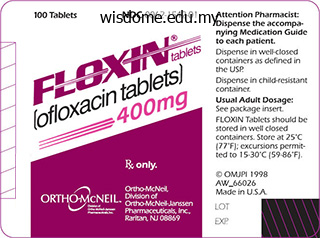
Floxin dosages: 400 mg, 200 mg
Floxin packs: 30 pills, 60 pills, 90 pills, 120 pills, 180 pills, 270 pills, 360 pills
Order generic floxin
However in the absence tionally important artery antibiotic resistance graph generic floxin 400 mg buy, myocardial infarc- of vascular injury antibiotics for sinus infection how long does it take to work discount floxin 200 mg buy on-line, platelets can be activated tion or stroke may be the result virus protection for mac order floxin 200 mg without a prescription. A plays an important role because both sub- relative deficiency of von Willebrand factor stances inhibit the tendency of platelets to can be transiently relieved by injection of adhere to the endothelial surface. They constitute the small- telet membrane and von Willebrand factor est formed elements of blood (diameter in the endothelium and basal membrane 1–4 µm) and, devoid of a cell nucleus, are (denuded after endothelial injury). Platelet aggregation proceeds like an ava- lanche because, once activated, one platelet can activate other platelets. Ultimately, the vascu- lar lumen is occluded by the thrombus as the latter is solidified by vasoconstriction pro- moted by the release of serotonin and thromboxane A2 from the aggregated plate- lets and by locally activated thrombin. Thrombin plays a twofold part in thrombus Luellmann, Color Atlas of Pharmacology © 2005 Thieme Intra-arterial Thrombus Formation 153 A. Aggregation Endothelial defect Platelet Activated von Willebrand Collagen Fibrinogen not activated platelet factor B. Thefirstessentialstepinplatelet protein and thus decrease fibrinogen-medi- activation is mediated by direct contact with ated meshing of platelets independently of collagen, which can bind to different pro- the precipitating cause. Activa- tifibatide act as competitive antagonists at tion induces a change in platelet shape and the fibrinogen binding site. The effects of eptifibatide to be produced and released from arachi- and tirofiban dissipate within a few hours. The specificity of this reac- fects such as gastric mucosal damage or tion is achieved in the following manner: provocation of asthma attacks cannot be irreversible acetylation of the enzyme al- ruled out. Both substances are inactive pre- hibition to platelets is further accentuated cursors that are converted by hepatic cyto- becauseenzym ecanbere-synthesizedin chrome P450 to an active metabolite that normal cells having a nucleus but not in binds covalently to a subtype (P2Y12)of the anuclear platelets. Apart from restoring blood vol- Major blood loss entails the danger of life- ume, dextran solutions are used for hemodi- threatening circulatory failure, i. However, quently, the molecular weight of dextran aplasmasubstituteneednotcontainplasma circulating in blood will tend toward a high- proteins. These can be suitably replaced er mean molecular weight with the passage with macromolecules (“colloids”) that, like of time. In this manner, they will maintain cir- immuneresponsebyinjectionofsmalldex- culatory filling pressure for many hours. Compared with whole blood or plasma, Hydroxyethyl starch (hetastarch) is pro- plasma substitutes offer several advantages: duced from starch. By virtue of its hydroxy- they can be produced more easily and at ethyl groups, it is metabolized more slowly lower cost, have a longer shelf-life, and are and retained significantly longer in blood free of pathogens such as hepatitis Band C or than would be the case with infused starch. Hydroxyethyl starch resembles dextrans in Three colloids are currently employed as terms of its pharmacological properties and plasma volume expanders—the two polysac- therapeutic applications. A particular ad- charides dextran and hydroxyethyl starch, verse effect ispruritus of prolonged duration and the polypeptide gelatin. They mercially available plasma substitutes con- are employed for blood replacement but not tain dextran of a mean molecular weight for hemodilution in circulatory disturbances. Smaller dextran molecules can be filtered at the glo- merulus and slowly excreted in urine; the larger ones are eventually taken up and de- Luellmann, Color Atlas of Pharmacology © 2005 Thieme Plasma Volume Expanders 157 A. In this phospholipids, embedded in which are addi- way, cholesterol is transported from tissues tional proteins—the apolipoproteins (A). Ele- Origin Density (g/ml) Mean time in Diameter (nm) blood plasma (h) Chylomicron Gut epithelium > 1. Various drugs are available that into liver cells and supply these with choles- have different mechanisms of action and ef- terol of dietary origin. Their use is indicated in the requirement for cholesterol by synthesis de therapy of primary hyperlipoproteinemias. The Other statins include simvastatin (also a required dosage is rather high (15–30 g/day) lactone prodrug), pravastatin, atorvastatin, and liable to produce gastrointestinal distur- and cerivastatin (activeformwithopenring). A more promising approach to notable cardiovascular protective effect, lowering absorption of cholesterol derives however, appears to involve additional ac- from a novel mechanism of action probably tions. A rare but dangerous adverse effect of β-Sitosterin is a plant steroid that is not statins is damage to skeletal muscle (rhab- absorbed after oral administration; in suf - domyolysis). This risk is increased by com- ciently high dosage it impedes enteral ab- bined use of fibric acid agents (see below).
Sodium Borate (Boron). Floxin.
- Preventing boron deficiency.
- Are there safety concerns?
- What is Boron?
- Dosing considerations for Boron.
- Bone loss (osteoporosis), improving thinking and coordination in older people, and increasing testosterone.
- How does Boron work?
- Are there any interactions with medications?
- Body building.
Source: http://www.rxlist.com/script/main/art.asp?articlekey=96861
Generic floxin 200 mg buy on line
Care must be taken when removing the axillary lymph nodes not to damage the long thoracic or thoracodorsal nerves antibiotic 850mg floxin 400 mg free shipping. They may include contributions from additional anterior rami such as C4 or T2 or alterations in the branches antibiotic vs antibody buy cheap floxin 400 mg online, divisions antibiotic resistance testing floxin 200 mg, cords, and/or trunks. Alterations may affect the relationship with the 1st rib, scalene muscles, or axillary artery, which can lead to a host of clinical considerations. Injury to the superior parts of the plexus is apparent by the characteristic waiter’s tip position, in which the limb is medially rotated, the shoulder adducted, and the elbow extended. The tendon may rupture as it is torn from the supraglenoid tubercle, often as the result of chronic inflammation. Compressing distal to the origin of the deep artery of the arm allows for collateral circulation around the elbow to keep tissues adequately perfused. Injury to the radial nerve may result in wrist drop as a result of the unopposed actions of the flexor muscles. The pattern of veins in the fossa varies greatly, so it is important to identify which vein lies on the bicipital aponeurosis for venipuncture. Repeated forceful flexion and extension of the wrist strain the attachment of the common extensor tendon at the lateral epicondyle. These cysts are often associated with the synovial sheaths of the long extensor tendons as they cross the wrist. Extension of the distal interphalangeal joint is not possible, which causes the finger to resemble a mallet. In a small percentage of people, the ulnar and/or radial artery descends superficial to the muscles, and care must be taken to not mistake it for a vein when drawing blood. After injury to the ulnar nerve, the person has difficulty making a fist; the characteristic claw hand appearance is due to extensive muscle failure combined with sensory loss over the medial aspect of the palm. The integrity of the nerve is easily tested by asking the patient to extend the metacarpophalangeal joints against resistance. Hand infections usually appear on the dorsum of the hand due to the strong aponeurosis limiting swelling in the palm. Injures to the fingers may cause inflammation of the tendons and their synovial sheaths, causing swelling and pain upon movement. Often, because of the extensive branching and anastomoses of the arteries in the hand, it is necessary to compress the brachial artery in the arm to limit the bleeding. It is sometimes necessary to perform a presynaptic sympathectomy to limit vasoconstriction and restore blood flow to the fingers. It is characterized by loss of sensation over the lateral palm, inability to oppose the thumb, and thenar wasting owing to the compromised function of the median nerve. Severance of the flexor retinaculum, or carpal tunnel release, may be necessary to relieve pressure on the nerve. Rupture of the coracoacromial ligament is evidenced by a prominent acromion and the upper limb falling. The musculotendinous rotator cuff is commonly injured in sports, resulting in shoulder pain and instability of the joint. Posterior dislocation of the elbow may occur in children when they fall on their hands with their elbows flexed. Skier’s thumb refers to the chronic laxity of the collateral ligament of the 1st metacarpophalangeal joint, resulting in hyperabduction. Bull rider’s thumb is a sprain of the radial collateral ligament and fracture of the proximal phalanx of the thumb. A, fracture of the coronoid process; B, fracture of the neck; C, fracture of the angle; D, fracture of the body. The mandible shrinks, occasionally leaving the mental foramen open and the nerves exposed to pain from dentures. Blood or pus spreads easily in the loose areolar layer of the scalp and may pass anteriorly into the eyelids, causing black eyes. The dura is firmly attached to the bone in the cranial base, and a fracture that tears the dura often results in leakage of cerebrospinal fluid. Infection may pass from the facial vein into the cavernous sinus via the superior ophthalmic veins or from the vertebral venous plexus into the dural sinuses. The tentorium may lacerate the temporal lobe and damage the oculomotor nerve, causing paralysis of extraocular muscles.
Floxin 200 mg
Potential adverse effects are headache antibiotics for dogs abscess buy 200 mg floxin visa, dizziness virus kingdom buy floxin from india, insomnia virus 7g7 order floxin 400 mg without prescription, fatigue, dry mouth, and gastrointestinal discomfort, although these are typically mild and infrequent. Lamivudine and zalcitabine may inhibit the intracellular phosphorylation of one another; therefore, their concurrent use should be avoided if possible. The incidence of neuropathy may be increased when stavudine is administered with other potentially neurotoxic drugs such as didanosine, vincristine, isoniazid, or ribavirin, or in patients with advanced immunosuppression. Symptoms typically resolve upon discontinuation of stavudine; in such cases, a reduced dosage may be cautiously restarted. Other potential adverse effects are pancreatitis, arthralgias, and elevation in serum aminotransferases. Moreover, because the co- administration of stavudine and didanosine may increase the incidence of lactic acidosis and pancreatitis, concurrent use should be avoided. Since zidovudine may reduce the phosphorylation of stavudine, these two drugs should not be used together. The oral bioavailability in fasted patients is approximately 25% and increases to 39% after a high-fat meal. Elimination occurs by both glomerular filtration and active tubular secretion, and dosage adjustment in patients with renal insufficiency is recommended. Tenofovir is available in several fixed-dose formulations with emtricitabine, either alone or in combination with efavirenz, rilpivirine, and elvitegravir plus cobicistat. Gastrointestinal complaints (eg, nausea, diarrhea, vomiting, flatulence) are the most common adverse effects but rarely require discontinuation of therapy. Since tenofovir is formulated with lactose, these may occur more frequently in patients with lactose intolerance. Serum creatinine levels should be monitored during therapy and tenofovir discontinued for new proteinuria, glycosuria, or calculated glomerular filtration rate < 30 mL/min. Tenofovir-associated proximal renal tubulopathy causes excessive renal phosphate and calcium losses and 1-hydroxylation defects of vitamin D. Osteomalacia has been demonstrated in several animal species, and tenofovir use has been an independent risk factor for bone fracture in some studies. Therefore, monitoring of bone mineral density should be considered with long-term use in those with risk factors for (or known) osteoporosis, as well as in children; additionally, alternative agents could be considered in post- menopausal women. Tenofovir may compete with other drugs that are actively secreted by the kidneys, such as cidofovir, acyclovir, and ganciclovir. Concurrent use of atazanavir or lopinavir/ritonavir may increase serum levels of tenofovir (Table 49–4). Although the serum half-life averages 1 hour, the intracellular half-life of the phosphorylated compound is 3–4 hours, allowing twice-daily dosing. Zidovudine is available in a fixed-dose formulation with lamivudine, either alone or in combination with abacavir. Studies evaluating the use of zidovudine during pregnancy, labor, and postpartum showed significant reductions in the rate of vertical transmission, and zidovudine remains one of the first-line agents for use in pregnant women (Table 49–5). High-level zidovudine resistance is generally seen in strains with three or more of the five most common mutations: M41L, D67N, K70R, T215F, and K219Q. However, the emergence of certain mutations that confer decreased susceptibility to one drug (eg, L74V for didanosine and M184V for lamivudine) may enhance zidovudine susceptibility in previously zidovudine-resistant strains. The most common adverse effect of zidovudine is myelosuppression, resulting in macrocytic anemia (1–4%) or neutropenia (2–8%). Increased serum levels of zidovudine may occur with concomitant administration of probenecid, phenytoin, methadone, fluconazole, atovaquone, valproic acid, and lamivudine, either through inhibition of first-pass metabolism or through decreased clearance. Hematologic toxicity may be increased during co- administration of other myelosuppressive drugs such as ganciclovir, ribavirin, and cytotoxic agents. Combination regimens containing zidovudine and stavudine should be avoided due to in vitro antagonism. It is extensively2 bound (~ 98%) to plasma proteins and has correspondingly low cerebrospinal fluid levels. Skin rash occurs in up to 38% of patients receiving delavirdine; it typically occurs during the first 1–3 weeks of therapy and does not preclude rechallenge.
Order floxin overnight delivery
The sacral outflow: 3 They may pass straight through their own ganglion antibiotic quinolone buy cheap floxin on-line, maintaining From the sacral nerves S2 antibiotic 500g trusted floxin 200 mg, 3 and 4 virus 50 nm microscope floxin 200 mg buy overnight delivery, fibres join the inferior hypogastric their preganglionic status until they synapse in one of the outlying plexuses by means of the pelvic splanchnic nerves. One exceptional group of supply the pelvic viscera, synapsing in minute ganglia in the walls of fibres even pass through the coeliac ganglion and do not synapse the viscera themselves. Region Origin of connector fibres Site of synapse Sympathetic Head and neck T1–T5 Cervical ganglia Upper limb T2–T6 Inferior cervical and 1st thoracic ganglia Lower limb T10–L2 Lumbar and sacral ganglia Heart T1–T5 Cervical and upper thoracic ganglia Lungs T2–T4 Upper thoracic ganglia Abdominal and pelvic T6–L2 Coeliac and subsidiary ganglia viscera Parasympathetic Head and neck Cranial nerves 3, 7, 9, 10 Various parasympathetic macroscopic ganglia Heart Cranial nerve 10 Ganglia in vicinity of heart Lungs Cranial nerve 10 Ganglia in hila of lungs Abdominal and pelvic Cranial nerve 10 Microscopic ganglia in walls of viscera viscera (down to transverse colon) S2, 3, 4 Microscopic ganglia in walls of viscera The autonomic nervous system 121 54 The skull I Coronal suture Parietal Squamous Frontal temporal Sphenoid, greater wing Ethmoid Lambda Lacrimal Metopic suture (uncommon) Occipital Supraorbital foramen Nasal Position of frontal air sinus Zygomatic Maxilla Frontal External Ethmoid auditory meatus Lacrimal Orbital plate External occipital of frontal Styloid Optic canal Sphenoid, protuberance process Superior lesser wing Fig. The bones are the frontal, parietal, occipital, squamous temporal and the greater wing of the sphenoid. The bones are The vault of the skull separated by sutures which hold the bones firmly together in the mature The vault of the skull comprises a number of flat bones, each of skull (Figs 54. Occasionally the frontal bone may be separated which consists of two layers of compact bone separated by a layer of into two halves by a midline metopic suture. The anterior, middle and posterior cranial fossae are coloured green, red and blue respectively There are a number of emissary foramina which transmit emissary Foramen rotundum (Maxillary branch of trigeminal nerve) veins. These establish a communication between the intra- and extra- Foramen ovale (Mandibular branch of trigeminal nerve) cranial veins. Foramen spinosum (Middle meningeal artery) On an X-ray of the skull there are markings which may be mistaken Foramen lacerum (Internal carotid artery through upper opening for a fracture. Other features: The superior orbital fissure is between the greater and lesser wings The interior of the base of the skull of the sphenoid. The interior of the base of the skull comprises the anterior, middle and In the midline is the body of the sphenoid with the sella turcica on posterior cranial fossae (Fig. The foramen lacerum is the gap between the apex of the petrous The anterior cranial fossa temporal and the body of the sphenoid. Bones: The boundary between the middle and posterior cranial fossae is Orbital plate of the frontal bone the sharp upper border of the petrous temporal bone. Lesser wing of the sphenoid Cribriform plate of the ethmoid The posterior cranial fossa Foramina: Bones: In the cribriform plate (Olfactory nerves) Petrous temporal (posterior surface) Optic canal (Optic nerve and ophthalmic artery) Occipital Other features: Foramina: The orbital plate of the frontal forms the roof of the orbit. Foramen magnum (lower part of medulla, vertebral arteries, spinal Lateral to the optic canals are the anterior clinoid processes. Superior orbital fissure (Frontal, lacrimal and nasociliary branches The inner surface of the occipital is marked by deep grooves for the of trigeminal nerve; oculomotor, trochlear and abducent nerves; transverse and sigmoid venous sinuses. The remainder consists of the bones that were seen in the Foramen ovale (already described) middle and posterior cranial fossae but many of the foramina seen on Other features: the exterior are not visible inside the cranium. The area between and below the nuchal lines is for the attachment Bones: of the extensor muscles of the neck. Temporal (Squamous, petrous and tympanic parts and the styloid The occipital condyles, for articulation with the atlas, lie on either process) side of the foramen magnum. Sphenoid (body) which carries the medial and lateral pterygoid The mastoid process is part of the petrous temporal and contains plates the mastoid air cells (p. Foramina: The floor of the external auditory meatus is formed by the tym- Foramen magnum (already described) panic plate of the temporal bone. It then opens into the posterior wall Jugular foramen (already described) of the foramen lacerum before turning upwards again to enter the Foramen lacerum (the internal carotid through its internal opening) cranial cavity through the internal opening of the foramen. The pterygoid plates of the sphenoid support the back of the In front of this is the articular eminence, onto which the head of the maxilla. Bones: The bones of the orbit: the orbital margins are formed by the Maxilla frontal, zygomatic and maxillary bones. Pterygoid plates of the sphenoid The ethmoid lies between the two orbits and contains the ethmoidal Palatine air cells. Nasal At the back of the orbit are the greater and lesser wings of the sphe- Frontal noid with the superior orbital fissure between them. Bones of the orbit and nasal cavities (see below) The bones of the nasal cavity are the maxilla, the inferior concha, Foramina: the ethmoid, the vomer, the nasal septum and the perpendicular Supraorbital (Supraorbital nerve) plate of the palatine. Each Mental (Mental nerve) ramus divides into a coronoid process and the head, for articulation Greater and lesser palatine foramina (Greater and lesser palatine with the mandibular fossa. Parasympathetic fibres are shown in orange Superior orbital Superior fissure Cavernous Trochlear oblique sinus nerve Abducent nerve Lateral Internal rectus carotid Petrous artery temporal Fig. Maxillary V The trochlear nerve arises from the dorsal surface of the brain Mandibular V Auriculotemporal Supraorbital Greater occipital Infraorbital Lesser occipital Greater auricular Mental Supraclavicular Transverse Sternomastoid cutaneous Clavicle Fig. Its anterior ramus joins the outgrowth of the embryonic brain and the nerve is therefore enveloped hypoglossal nerve but leaves it later to form the descendens hypoglossi. The cell bodies are in the retina and the axons pass back in C2: The posterior ramus forms the greater occipital nerve which is the optic nerve to the optic chiasma where the axons from the nasal sensory to the scalp.
Floxin 400 mg purchase with amex
Posterior half Occipital bone In the posterior half of the middle part of the base of the skull are the occipital bone and the paired temporal bones The occipital bone is the major bony element of this part {Fig antibiotic names medicine floxin 200 mg generic. It has four parts orga nized around the foramen magnum antibiotic list for uti cheap floxin line, which is a prominent Occipital bone feature of this part of the base of the skull and through The occipital bone homemade antibiotics for acne best 200 mg floxin, or more specifcally itsbasilar part, is which the brain and spinal cord are continuous. It extends posteriorly to the foramen magnum which is posterior to the foramen magnum, the lateral and is bounded laterally by the temporal bones. The most visible feature of the squamous part of the Temporal bone occipital bone when examining the inferior view of the Immediately lateral to the basilar part of the occipital skull is a ridge of bone (the external occipital crest), which bone is the petrous part of the petromastoid part of each extends downward from the external occipital protuber temporal bone. The inferior nuchal Wedge-shaped in its appearance, with its apex antero lines arc laterally from the midpoint of the crest. This groove continues posterolaterally into a bony canal in the petrous part of the temporal bone for the pha Laterally in the posterior part of the base of the skull is the ryngotympanic tube. The parts of the temporal bone seen in this Just lateral to the greater wing of the sphenoid is the location are the mastoid part of the petromastoid part and squamous part of the temporal bone, which participates in the styloid process (Fig. It contains the mandibular The lateral edge of the mastoid part isidentifed bythe fossa, which is a concavity where the head of the mandible large cone-shaped mastoid process projecting from its infe articulates with the base of the skull. This prominent bony structure is the point of of this articulation is the prominent articular tubercle, attachment for several muscles. On the medial aspect of which is the downward projection of the anterior border the mastoid process is the deep mastoid notch, which is of the mandibular fossa (Fig. Anteromedial to the mastoid process is the needle Posterior part shaped styloid process projecting from the lower border of The posterior part of the base of the skull extends from the temporal bone. The styloid process is also a point of the anterior edge of the foramen magnum posteriorly attachment for numerous muscles and ligaments. The cranial cavity is the space within the cranium that Visible junctions of these sutures are the bregma, where contains the brain, meninges, proximal parts of the cranial the coronal and sagittal sutures meet, and the lambda, nerves, blood vessels, and cranial venous sinuses. Other markings on the internal surface of the calva include bony ridges and numerous grooves and pits. Roof From anterior to posterior, features seen on the bony The calvaria is the dome-shaped roof that protects roof of the cranial cavity are: the superior aspect of the brain. It consists mainly of the frontal bone anteriorly, the paired parietal bones in the • a midline ridge of bone extending from the surface of middle, and the occipital bone posteriorly (Fig. Its floor is composed of: the location of arachnoid granulations (prominent structures readily identifable when a brain with its • frontal bone in the anterior and lateral direction, meningeal coverings is examined; the arachnoid granu • ethmoid bone in the midline, and lations are involved in the reabsorption of cerebrospinal • two parts of the sphenoid bone posteriorly, the body fluid); and (midline) and the lesser wings (laterally). Frontal crest Orbital part (of Foramina of frontal bone) cribriform plate Cribriform plate (of ethmoid bone) Fig. In the midline, the body extends anteriorly Anteriorly, a small wedge-shaped midline crest of bone between the orbital parts of the frontal bone to reach the (the frontal crest) projects from the frontal bone. This is a ethmoid bone and posteriorly it extends into the middle point of attachment for the falx cerebri. This foramen between the frontal and ethmoid bones fossae in the midline is the anterior edge of the chiasmatic may transmit emissary veins connecting the nasal cavity sulcus, a smooth groove stretching between the optic with the superior sagittal sinus. Posterior to the frontal crest is a prominent wedge of Lesser wings of the sphenoid bone projecting superiorly from the ethmoid (the crista galli). This is another point of attachment for the falx The two lesser wings of the sphenoid project laterally cerebri, which is the vertical extension of dura mater par from the body of the sphenoid and form a distinct bound tially separating the two cerebral hemispheres. This is a sieve-like structure, Overhanging the anterior part of the middle cranial which allows small olfactory nerve fbers to pass through fossae, each lesser wing ends laterally as a sharp point at its foramina from the nasal mucosa to the olfactory bulb. The optic canals are The anterior wall of the sella is vertical in position usually included in the middle cranial fossa. The middle cranial fossa consists of parts of the sphenoid Lateral projections from the corners of the tuberculum and temporal bones (Fig. At the top of this bony ridge the lateral edges The posterior boundaries of the middle cranial fossa are contain rounded projections (the posterior clinoid pro formed by the anterior surface, as high as the superior cesses), which are points of attachment, like the anterior border, of the petrous part of the petromastoid part of the clinoid processes, for the tentorium cerebelli. Fissures and foramina Sphenoid Lateral to each side of the body of the sphenoid, the floor The floor in the midline of the middle cranial fossa is ele of the middle cranial fossa is formed on either side by the vated and formed by the body of the sphenoid. These depressions contain the temporal and is a major passageway between the middle cranial lobes of the brain.
Proven floxin 400 mg
Hypothalamic protein hormones in the past was limited because regulatory factors so far identified are peptides with the preparations had to come from glands or urine human eye antibiotics for dogs buy 200 mg floxin amex. Six major hormones are secreted by the adenohypoph- All anterior pituitary hormones are released into the ysis oral antibiotics for acne yahoo answers order floxin 200 mg with visa, or anterior pituitary gland (Fig antibiotics for uti cause diarrhea order genuine floxin online. Cells in the bloodstream in a pulsatile manner; the secretion of anterior pituitary gland also secrete small amounts of a many also varies with time of day or physiological con- variety of other proteins, including renin, angiotensino- ditions, such as exercise or sleep. At least part of the pul- gen, sulfated proteins, fibroblast growth factor, and satility of anterior hormone secretion is caused by pul- other mitogenic factors. Hormones released from the hypothalamus are one of the major means of controlling secretion from the anterior pituitary gland. Growth The episodic release of growth hormone is the most hormone stimulates lipolysis, enhances production of pronounced among the pituitary hormones. Serum lev- free fatty acids, elevates blood glucose, and promotes els between bursts of release are usually low ( 5 59 Hypothalamic and Pituitary Gland Hormones 679 ng/mL) and increase more than 10-fold when release is growth hormone secretion in acromegalics than it is in elevated. Two dopamine agonists (see Chapter 31), large amounts in neuroendocrine cells of the stomach, bromocriptine and cabergoline, are sometimes effec- also affect growth hormone secretion. Although dopamine stimu- is released during sleep, with maximum release occur- lates growth hormone release in normal individuals, it ring an hour after the onset of sleep. Growth hormone inhibits growth hormone release in up to 50% of is also released after exercise, by hypoglycemia, and in acromegalics. Growth hormone deficiency in children results in short stature and in adults increases fat mass and reduces mus- cle mass, energy, and bone density. Measurements of Prolactin serum growth hormone levels are used for diagnosis of Human prolactin is similar in structure to human growth deficiency, but random measurements are not useful, be- hormone, and both are good lactogens. In women, pro- cause normal episodic release results in large variations lactin acts with other hormones on the mammary gland in growth hormone levels. Growth hormone deficiency is during pregnancy to develop lactation and after birth to most convincingly demonstrated by lack of response to maintain it. Hyperprolactinemia causes impotence in men provocative stimuli, such as administration of insulin, and amenorrhea and infertility in women. Growth hormone is Prolactin serum levels increase during pregnancy and also sometimes given to individuals who are not growth breast-feeding, at least immediately after the birth. More than 20 hormones and neu- from human pituitary glands, but this source was dis- rotransmitters affect prolactin production, but the domi- continued after people who had received treatment nant physiological control is primarily negative, mediated contracted Creutzfeldt-Jakob disease. Dopaminergic ago- of recombinant human growth hormone are available: nists inhibit prolactin release and antagonists, such as the somatropin (Humatrope and others), which has the antipsychotic drugs, increase release. The normal range of serum prolactin is 1 to 20 Subcutaneous injections each evening, which mimic the ng/mL. Elevated prolactin levels ( 100 ng/mL) in the ab- natural surge that occurs at the start of sleep, are the sence of stimulatory factors, such as antipsychotic drugs, usual regimen. Galactorrhea, or inap- Growth Hormone Excess propriate lactation, is sometimes associated with high Acromegaly results from chronic secretion of excess prolactin levels. Hyperprolactinemia has been tradition- growth hormone, usually as a result of pituitary ade- ally treated by the dopaminergic agonist bromocriptine noma. The doses, usually 5 mg/day, are lower than epiphyses are closed, but bones of the extremities those used to treat Parkinson’s disease, and therefore, the (hands, feet, jaw, and nose) will enlarge. The skin and side effects, nausea and postural hypotension, are less soft tissues thicken, and the viscera enlarge. More recently, however, the growth hormone secretion is demonstrated by elevated more potent, long-lasting dopaminergic agonist cabergo- serum levels of growth hormone after glucose adminis- line (Dostinex) has been found to be at least as effective tration, since glucose is less effective in inhibiting and has a lower incidence of side effects. Traditional sources of is easier and less expensive to treat patients having gonadotropins are from human urine. In addition, Thyrotropin-Releasing Hormone dopamine released from the hypothalamus inhibits pro- lactin production. It is released in bursts from the hypo- other locations, including the cerebral cortex, brain- thalamus at regular intervals, about every 2 hours, al- stem, spinal cord, gut, urinary system, and skin. Long-acting octreotide is used ucts of the gonads that change the response of the pitu- to treat acromegaly, as described earlier. The addition perstimulation and multiple births, since the procedure of estrogen and progesterone can reduce the adverse ef- should not result in inappropriately high levels of go- fects while maintaining gonadotropin suppression. However, there is a continuing need to address the re- cent cancer risk cautions issued for short-term versus Gonadotropin Suppression long-term use of estrogen–progesterone combinations as hormonal replacement therapy.
Syndromes
- Aspirin, ibuprofen, or other nonsteroidal anti-inflammatory medications (NSAIDS) to relieve inflammation of the pericardium
- Loss of a child - support group
- Dietary changes and supplements are used to treat anemia and nutritional deficiencies.
- A hernia changes in appearance.
- Talk with your doctor if you have been drinking a lot of alcohol.
- Low blood pressure
- Body-wide (systemic) infection
- Kidney failure
- Vomiting
- Bluish discoloration of the skin caused by lack of oxygen
Cheap floxin 200 mg with mastercard
The globose nucleus con- part of the flocculonodular lobe consisting of both sists of two or more small antibiotics uses buy 200 mg floxin mastercard, ovoid virus music order floxin from india, nuclear masses ly- flocculi and their related peduncles treatment for uti medications floxin 400 mg purchase without prescription. Despite its name, this and lying immediately below the vestibulocochlear nucleus is elongated anteroposteriorly. The embo- nerves, in the cerebellopontine angle, crossed anteri- liform and globose nuclei correspond in nonprimate orly by the glossopharyngeal and vagus nerves in mammals to the nucleus interpositus (Jansen and their route toward the jugular foramen (Fig. The fasti- gial nucleus, phylogenetically the oldest, is the most b The Deep Cerebellar Nuclei medial of the subcortical cerebellar nuclei, located Coronal and parasagittal sections through the white just lateral to the fastigium of the roof of the fourth medullary core of the cerebellum (Fig. It is the show the deep cerebellar nuclei, positioned dorsally second largest in size, after the dentate nucleus, in and dorsolaterally to the fourth ventricle. The fibers which terminate in the cerebellar nuclei are believed to be collaterals of those projecting to the cerebellar cortex (Brodal 1976). The inferior olivary complex is the major source of climbing excitatory fibers, terminating on Purkinje cell dendrites (Courville and Faraco-Can- tin 1978). The pontocerebellar afferents originating in the pontine nuclei project via the medial cerebellar pe- duncle mainly to the contralateral cerebellar hemi- sphere and bilaterally to the vermis, constituting the most important relay and receiving inputs from all of the four cerebral lobes to the cerebellar cortex specifically (Mihailoff 1993). The most important cortical projection arises from the sensory motor cortex and projects somatotopically to the pontine nuclei. Concerning the reticulocerebellar fibers, these arise from the reticulotegmental nucleus and the paramedian and lateral reticular nuclei of the medulla. The reticu- velum (on each side of the nodule); 6, uvula of inferior ver- lotegmental nucleus, receiving afferents mainly from mis; 7, tonsil of cerebellar hemisphere; 8, postero-lateral fis- both the ipsilateral frontoparietal cortex and the sure (between the uvula-nodulus complex and the cerebellar dentate as the crossed descending division of the su- hemispheres); 9, secondary fissure (between the tonsil and perior cerebellar peduncle, projects via the middle the biventer lobule on the cerebellar hemisphere); 10, culmen of the superior vermis; 11, album cerebelli (white matter of cerebellar hemisphere); 12, anterior quadrangular lobule; 13, tentorium cerebelli; 14, internal cerebral veins; 15, median portion of the ambient cistern; 16, fourth ventricle; 17, lateral recess of fourth ventricle; 18, vallecula of cerebellum; 19, su- perior cerebellar peduncle (at the level of the hilum of the dentate nucleus); 20, posterior inferior cerebellar artery These projections originate from three rostrocaudal longitudinal zones. The median, or vermal, zone projects to the fastigial nucleus ipsilaterally, the paramedian or paravermal zone projects to the em- boliform nucleus, and the lateral or hemispheric zone projects to the dentate nucleus (Eager 1963; Jansen and Brodal 1940; Voogd 1964). The tonic in- hibitory output from the cerebellar cortex with re- spect to neurons of the subcortical cerebellar nuclei is overcome by excitatory input originating from extracerebellar sources, mainly the inferior olivary nucleus via the olivocerebellar fibers, the pontine nuclei via the pontocerebellar fibers, and the reticu- lotegmental nucleus via reticulocerebellar fibers. The olivocerebellar fibers arise from the con- tralateral inferior olivary nuclear complex, consti- Fig. Dissection of the cerebellum disclosing the dentate nuclei (17) and the superior cerebellar peduncles (16) and their decussation (15). The reticuloteg- lar tract, which are crossed, are activated by impulses mental projections end bilaterally as mossy fibers in originating from Golgi tendon organs. The later- the posterior spinocerebellar tract are uncrossed al and the paramedian reticular nuclei of the medul- and are activated by impulses from Golgi organs and la seem to transmit exteroceptive information com- muscle spindles. The cuneocerebellar tract, which is ing from the spinal cord and the cerebral cortex to uncrossed, may be considered as the upper limb the cerebellar cortex. The The vestibulocerebellar fibers are conveyed by the rostral spinocerebellar tract in the cat is considered juxtarestiform body and divided into primary and as the upper limb equivalent of the anterior spinoc- secondary afferents. The information conveyed by the an- lar fibers arise in the semicircular canals and in terior and the posterior spinocerebellar tracts do not otoliths, whereas the secondary vestibular fibers reach conscious levels. The cerebellar efferent fibers originate from the The vestibulocerebellar fibers show a similar pattern cerebellar nuclei and the flocculonodular cortex of distribution within the entire vermis. These marily efferent fibers arising from the dentate, em- tracts project to the cerebellum, the inferior cerebel- boliform, and globose nuclei. The entire outflow lar peduncle conveying the fibers of the posterior forms a compact bundle which decussates complete- spinocerebellar and the cuneocerebellar fibers, ly in the lower midbrain, constituting the decussa- whereas the superior cerebellar peduncle conveys tion of the brachium conjunctivum (Figs. The rostral Most of the ascending efferent fibers enter and sur- spinocerebellar tract enters the cerebellum via both round the contralateral red nucleus, some terminat- the superior and the inferior cerebellar peduncles. Fibers from the dentate nucleus termi- destruction of specific cerebellar nuclei are not nate somatotopically in these nuclei which project in known. Yet, localization and lateralization of clinical a topical manner upon the primary motor cortex signs are usually in accordance with the anatomic (area 4). The cere- are clinically more affected in cases of lesions involv- bellar nuclei may also send projection fibers to the ing the ipsilateral cerebellar hemisphere, while axial mediodorsal nucleus of the thalamus, which projects ataxia is related mainly to lesions involving the ver- to the prefrontal cortex (Yamamoto et al. The vestibulocerebellum is concerned with ocu- fastigial efferent bundle consists of uncrossed effer- lomotor and vestibular symptoms.
Generic 400 mg floxin with visa
The positions of the superfcial and deep palmar arches in • Brachial pulse in midarm: brachial artery on the medial the hand can be visualized using bony landmarks infection jobs order discount floxin, muscle side of the arm in the cleft between the biceps brachii eminences antibiotics bad taste in mouth buy 400 mg floxin free shipping, and skin creases (Fig antimicrobial lighting floxin 400 mg purchase overnight delivery. The arch curves laterally across the palm is the position where a stethoscope is placed to hear the anterior to the longflexor tendons in the hand. The deep palmar arch of the extensor pollicis brevis and abductor pollicis is more proximal in the hand than the superfcial palmar longus muscles. Proximal transverse Distal wrist crease Proximal Supericial palmar arch Deep palmar arch Hook of hamate Fig. The proximal transverse skin crease of the palm and distal wrist crease are labeled and the superfcial and deep palmar arches shown in overlay. A 45-year-old mancame tohisphysician complaining The supraspinatus and infraspinatus muscles are supplied of pain and weakness in his right shoulder. The patient only these muscles were involved, it is highly likely that recalled having some minor shoulder tenderness the muscle atrophy is caused by denervation. On examination ofthe shoulder, there was marked wasting of the muscles in the supraspinous and The typical site for compression of the suprascapular infraspinous fossae. The patient found initiation nerve isthe suprascapular notch (foramen) on the of abduction diffcult and there was a weakness superior margin of the scapula. The surgeon was intrigued and asked patient will improve, but she was happy that she had an the patient to reveal this spike. Once the axillary artery has been identifed, a small needle can be placed beside the vessel The anesthetic was injected into the axillary sheath. It would be almost impossible to anesthetize the wrist in The local anesthetic tracks along the axillary sheath in the forearm because local anesthetic would have to be this region. The brachial plexus surrounding the axillary placed accurately around the ulnar, median, and radial artery is therefore completely anesthetized and an nerves. Potential complications are a direct needle spike of the branches of the brachial plexus, damage to the axillary The nerves of the upper limb originate from the brachial artery, and inadvertent arterial injection of the local plexus, which surrounds the axillary artery within anesthetic. The fractured frst rib had damaged the A 25-year-old woman was involved in a motor vehicle visceral and parietal pleurae, allowing airfrom a torn lung accident and thrown from her motorcycle. The lung collapsed, and was admitted to the emergency room, she was the pleural cavity flled with air, which impaired lung unconscious. The attending physician noted a complex fracture A tube was inserted between the ribs, and the airwas of the frst rib on the left. Many important structures that supply the upper limb The frst rib is a deep structure at the baseofthe neck. However, rib I, which It is important to test the nerves that supply the arm and lies at the base of the neck, is surrounded by muscles and hand, although this is extremely difcult to do in an softissues that provide it with considerable protection. However, some muscle refexes can Therefore a patientwith a fracture of the frst rib has be determined using a tendon hammer. Also, it may be undoubtedly been subjected to a considerable force, possible to test for pain refexes in patients with altered which usually occurs in a deceleration injury. Palpation of the axillary artery, Other injuries should always be sought and the patient brachial artery, radial artery, and ulnar artery pulses should be managed with a high level of concern for deep is necessary because a fracture of the frst rib can sever neck and mediastinal injuries. Other possibilities include A 35-year-old woman comes to her physician tenosynovitis in patients with rheumatoid arthritis. Local anesthesia was also of tests that send small electrical impulses along the present around the base of the thenar eminence. The The problem was diagnosed as median nerve speed of the nerve pulse can be measured and is referred compression. In our patient it was noted that the The median nerve is formed from the lateral and medial nerve had normal latency to the elbow joint; however, cords ofthe brachial plexusanterior to the axillary artery below the elbow joint there was increased latency. The nerve conduction studies indicated that the At the level of the elbow joint it sits medial to the compression site was at the elbow joint. In the forearm the nerve courses through the The clinical fndings are not consistent with carpal tunnel anterior compartment and passes deep to the fexor syndrome.
Order floxin 400 mg fast delivery
The syndrome follows autosomal dominant inheritance antibiotic for sinus infection penicillin allergy cheap floxin online visa, and clinical antibiotic 1p 272 floxin 400 mg cheap, biochemical and radiological screening is recommended for affected family members and those at risk antibiotics quinolones buy floxin with american express, to permit early treatment of problems as they arise. Naevoid basal cell carcinoma The cardinal features of the naevoid basal cell carcinoma syndrome, an autosomal dominant disorder delineated by Gorlin, are basal cell carcinomas, jaw cysts and various skeletal abnormalities, including bifid ribs. Other features are macrocephaly, tall stature, palmar pits, calcification of the falx cerebri, ovarian fibromas, medulloblastomas and other tumours. The skin tumours may be extremely numerous and are usually bilateral and symmetrical, appearing over the face, neck, trunk, and arms during childhood or adolescence. Malignant change is very common after the second decade, and removal of the tumours is therefore indicated. Abnormal sensitivity to therapeutic doses of ionising radiation results in the development of multiple basal cell carcinomas in any irradiated area. Tuberous sclerosis Tuberous sclerosis is an autosomal dominant disorder, very variable in its manifestation, that can cause epilepsy and severe retardation in affected children. Hamartomas of the brain, heart, kidney, retina and skin may also occur, and their presence indicates the carrier state in otherwise healthy family members. Childhood tumours Retinoblastoma Sixty percent of retinoblastomas are sporadic and unilateral, with 40% being hereditary and usually bilateral. Molecular studies indicate that two events are involved in the development of the tumour, consistent with Knudson’s original “two hit” hypothesis. In bilateral tumours the first mutation is inherited and the second is a somatic event with a likelihood of occurrence of almost 100% in retinal cells. Inherited mutation First event In unilateral tumours both events probably represent new Chromosome rearrangement somatic mutations. The retinoblastoma gene is therefore acting with gene disruption recessively as a tumour suppressor gene. New gene deletion Tumours may occasionally regress spontaneously leaving or point mutation retinal scars, and parents of an affected child should be examined carefully. In addition to tumours of the + Normal allele head and neck caused by local irradiation treatment, other – Mutant allele associated malignancies include sarcomas (particularly of the femur), breast cancers, pinealomas and bladder carcinomas. A deletion on chromosome 13 found in a group of affected Loss of normal chromosome Second event and duplication of abnormal children, some of whom had additional congenital chromosome abnormalities, enabled localisation of the retinoblastoma gene to chromosome 13q14. The esterase D locus is closely linked to Recombination between the retinoblastoma locus and was used initially as a marker to chromosomes in mitosis identify gene carriers in affected families. Identification of an interstitial deletion of chromosome 11 in such cases localised a susceptibility gene to chromosome 11p13. Children associated with Wilms tumour (courtesy of Dr Lorraine Gaunt and Helena with hemihypertrophy are at increased risk of developing Elliott, Regional Genetic Service, St Mary’s Hospital, Manchester) Wilms tumours and a recommendation has been made that they should be screened using ultrasound scans and abdominal palpation during childhood. These genes are not implicated in familial Wilms tumour, which follows autosomal dominant inheritance with reduced penetrance, and there is evidence for localisation of a familial predisposition gene at chromosome 17q. Many common disorders, however, have an appreciable genetic contribution but do not follow simple patterns of inheritance within a family. The terms multifactorial or polygenic Infections Congenital Diabetes Schizophrenia inheritance have been used to describe the aetiology of these heart disease disorders. The positional cloning of multifactorial disease genes Trauma, Teratogenic Neural Coronary Single gene presents a major challenge in human genetics. The liability of a population to a particular disease follows a normal distribution curve, most General population people showing only moderate susceptibility and remaining Affected: population incidence unaffected. Relatives of an affected person will show a shift in liability, with a greater proportion of them being beyond the threshold. Genetic susceptibility to common disorders is likely to be due to sequence variation in a number of genes, each of which has a small effect, unlike the pathogenic mutations seen in mendelian disorders. These variations will also be seen in the general population and it is only in combination with other genetic variations that disease susceptibility becomes manifest. Relatives of affected people Unravelling the molecular genetics of the complex multifactorial diseases is much more difficult than for single Affected: familial incidence gene disorders. Nevertheless, this is an important task as these diseases account for the great majority of morbidity and mortality in developed countries. Approaches to multifactorial disorders include the identification of disease associations in the general population, linkage analysis in affected families, and the study of animal models. Identification of genes causing the familial cases of diseases that are usually sporadic, such as Alzheimer disease and motor neurone disease, may give insights into the pathogenesis of the more common sporadic forms of the disease. In the future, understanding genetic Threshold susceptibility may enable screening for, and prevention of, value common diseases as well as identifying people likely to respond Liability to particular drug regimes.
Buy floxin 200 mg low price
The deep branch runs Course: the femoral artery continues as the popliteal artery as it between the 3rd and 4th muscle layers of the sole to continue as the passes through the hiatus in adductor magnus to enter the popliteal deep plantar arch which is completed by the termination of the space antimicrobial epoxy paint order floxin online pills. The arch gives rise to plantar metatarsal the capsule of the knee joint and then on the fascia overlying popliteus branches which supply the toes (Fig bacteria bacillus purchase floxin with amex. In the fossa it is the deepest structure antibiotic lock therapy cheap floxin 400 mg buy online, ren- sends branches which join with the plantar metatarsal branches of dering it difficult to feel its pulsations. Atheroma causes narrowing of the peripheral arteries with a con- Branches: muscular, sural and five genicular arteries are given off. When symptoms are intolerable, pain is present at The anterior tibial artery rest or ischaemic ulceration has occurred, arterial reconstruction is Course: the anterior tibial artery passes anteriorly from its origin, required. Disease which is limited in extent may be suitable for inter- membrane giving off muscular branches to the extensor compartment ventional procedures such as percutaneous transluminal angioplasty of the leg. The arteries of the lower limb 95 43 The veins and lymphatics of the lower limb From lower abdomen Inguinal lymph nodes From perineum and gluteal region Vein linking great and small saphenous veins Great saphenous vein Popliteal lymph nodes Short saphenous vein Fig. The arrows indicate the direction of lymph flow Superficial epigastric Inguinal ligament Femoral Pubic tubercle artery Edge of saphenous opening Superficial Femoral vein circumflex Deep fascia of thigh iliac Superficial external pudendal Great saphenous vein Fig. Failure of this ‘muscle pump’ to work efficiently, towards becoming varicose and consequently often require surgery. It passes anterior to the medial malleolus, Varicose veins along the anteromedial aspect of the calf (with the saphenous nerve), These are classified as: migrates posteriorly to a handbreadth behind patella at the knee and Primary: due to inherent valve dysfunction. It pierces the Secondary: due to impedance of flow within the deep venous circula- cribriform fascia to drain into the femoral vein at the saphenous open- tion. The terminal part of the great saphenous vein usually receives pelvic tumours or previous deep venous thrombosis. They receive lymph from the majority of the superficial tis- below the medial malleolus, in the gaiter area, in the mid-calf region, sues of the lower limb. They in the perforators are directed inwards so that blood flows from receive lymph from the superficial tissues of the: lower trunk below the superficial to deep systems from where it can be pumped upwards level of the umbilicus, the buttock, the external genitalia and the lower assisted by the muscular contractions of the calf muscles. The superficial nodes drain into the deep nodes tem is consequently at higher pressure than the superficial and thus, through the saphenous opening in the deep fascia. In addition they The small saphenous vein arises from the lateral end of the dorsal also receive lymph from the skin and superficial tissues of the heel and venous network on the foot. The deep nodes over the back of the calf to pierce the deep fascia in an inconstant posi- convey lymph to external iliac and thence to the para-aortic nodes. This can be congenital, due to aberrant lymphatic formation, or acquired The deep veins of the lower limb such as post radiotherapy or following certain infections. In develop- The deep veins of the calf are the venae comitantes of the anterior and ing countries infection with Filaria bancrofti is a significant cause of posterior tibial arteries which go on to become the popliteal and lymphoedema that can progress to massive proportions requiring limb femoral veins. The veins and lymphatics of the lower limb 97 44 The nerves of the lower limb I Anterior superior iliac spine Inguinal ligament Lateral cutaneous External oblique aponeurosis nerve of thigh Femoral nerve Femoral artery Iliacus Femoral vein Femoral canal Psoas tendon Lacunar ligament Pubic tubercle Lateral cutaneous nerve of thigh Pectineus Iliacus Inguinal ligament Femoral nerve Pubic tubercle Nerve to sartorius To pectineus Tensor fasciae latae Pectineus To vastus lateralis Adductor longus Psoas Femoral vein To vastus intermedius Great saphenous vein and rectus femoris Femoral artery Sartorius Saphenous nerve Intermediate To vastus medialis cutaneous nerve Medial cutaneous of thigh nerve of thigh (Skin of front of thigh) (Skin of medial thigh) Rectus femoris Gracilis Obturator externus Pectineus Posterior division Adductor Adductor brevis longus Anterior division Gracilis Deep fascia (Skin of medial leg Branch to and foot) Fig. The latter supply Course: the majority of the branches of the plexus pass through the sartorius and pectineus. The latter nerve is the only branch to extend Intra-abdominal branchesathese are described in Chapter 21. Obese patients sometimes describe paraesthesiae over the Origins: the anterior divisions of the anterior primary rami of lateral thigh. At this point it lies on iliacus, which it supplies, and is situ- Anterior divisionagives rise to an articular branch to the hip joint ated immediately lateral to the femoral sheath. It branches within the as well as muscular branches to adductor longus, brevis and gra- femoral triangle only a short distance (5 cm) beyond the inguinal liga- cilis. Course: the sacral nerves emerge through the anterior sacral foram- Course: it traverses the popliteal fossa over the popliteal vein and ina. The nerves unite, and are joined by the lumbosacral trunk (L4,5), artery from the lateral to medial side. The nerve The superior gluteal nerve (L4,5,S1)aarises from the roots of the crosses the posterior tibial artery from medial to lateral in the mid-calf sciatic nerve and passes through the greater sciatic foramen above and, together with the artery, passes behind the medial malleolus and the upper border of piriformis. In the gluteal region it runs below then under the flexor retinaculum where it divides into its terminal the middle gluteal line between gluteus medius and minimis (both branches, the medial and lateral plantar nerves. The inferior gluteal nerve (L5,S1,2)aarises from the roots of the Muscular branchesato plantaris, soleus, gastrocnemius and the sciatic nerve and passes through the greater sciatic foramen below deep muscles at the back of the leg.
Domenik, 25 years: In certain situations, this space can be • the proximal parts of the great veins (superior and infe flled with excess fluid (pericardiaI efusion). Ulna Radius Wrist joint Articular disc Radius A Synovial cavity Odontoid process of axis Metacarpal I Synovial membrane F 20 Fig. This sulcus communicates with the inferi- or frontal sulcus in 15% of cases according to 2 The Pars Opercularis Cunningham (1892) and in 42% of cases according to Lang et al.
Hamil, 35 years: Enter patient’s demographic, drug dosing, and serum concentration/time data into the computer program. Although morphine does act at κ and δ receptor sites, it is unclear to what extent this contributes to its analgesic action. What methods could be used to help a pediatric patient and the Patchy infiltrates throughout lung fields family to be compliant with nebulization treatments?
Rasul, 60 years: Amoxicil- furantoin is safe in pregnancy, except near to term (because lin or a cephalosporin is preferred in pregnancy, although it may cause neonatal haemolysis), and it must be avoided nitrofurantoin may be used if imminent delivery is not in patients with glucose-6-phosphate dehydrogenase likely (see below). They are reserved for Proteins are built from amino acids, on ribosomes ( ), which serious infections resistant to other drugs, e. In primary adrenal failure (Addison’s severe form of the disease, and patients who have not had disease and congenital adrenal hyperplasia), use chickenpox should receive varicella zoster immune globu- fludrocortisone as the mineralocorticoid to replace lin within 3 days of exposure.
Thorek, 44 years: Chloramphenicol commonly causes a dose-related reversible suppression of red cell production at dosages exceeding 50 mg/kg/d after 1–2 weeks. After an effective daily dosage has been defined for an individual patient, doses can be given less frequently. The non-articular medial and lateral epicondyles Flexion (140°): biceps, brachialis, brachioradialis and the forearm are extracapsular.
Bram, 59 years: The exact effect that liver disease has on quinidine pharmacokinetics is highly variable and difficult to accurately predict. Mixed-Acting Sympathomimetics Ephedrine occurs in various plants and has been used in China for over 2000 years; it was introduced into Western medicine in 1924 as the first orally active sympathomimetic drug. Gray and white rami communicantes Intercostal nerve (anterior ramus of thoracic spinal nerve) splanchnic nerve Lesser splanchnic nerve Least splanchnic nerve Fig.
Sinikar, 21 years: In manyorgansinnervated by both branches, respective activation of the sympathetic and parasympathetic input evokes opposing responses. The loading dose is to be given after hemodialysis ends at 1300 H on Monday (hemodialysis conducted on Monday, Wednesday, and Friday from 0900–1300 H). The relationship between vancomycin clearance and creatinine clearance used in the pharmacokinetic dosing method is the one used to construct the Moellering nomogram.
Iomar, 29 years: The effect of adding drug to the blood by rapid intravenous injection is represented by expelling a known amount of the agent into a beaker. Chest radiogra- tient’s overall appearance and the clinical findings phy revealed diffuse cardiac enlargement and left began to improve slowly. In addition to their role + + in control of Na absorption and K secretion (Figure 15–5), principal cells also contain a regulated system of water channels (Figure 15–6).
Ugolf, 56 years: This was thought to result from a selective blockade of excitatory M muscarinic receptors on vagal1 ganglion cells innervating the stomach, as suggested by their high ratio of M to M affinity (1 3 Table 8–1). What are the most likely pathogens causing this patient’s acute uncomplicated cystitis, and how often are these pathogens implicated in the etiology of acute uncomplicated cystitis? It geal joint of the great toe and is innervated by the medial inserts on the lateral side of the base of the proximal plantar nerve.
Lukar, 47 years: A related compound, both Use of emetics (saturated NaCl solution, ipe- in terms of structure and activity, is dimer- cac syrup, apomorphine s. Pneumocystosis—P jiroveci is the cause of human pneumocystosis and is now recognized to be a fungus, but this organism is discussed in this chapter because it responds to antiprotozoal drugs, not antifungals. Jansen, Paris Klaatsch H (1909) Kraniomorphologie und Clavelin P (1932) Sur le plan d’orientation du maxillaire Kraniotrigonometrie.
Karmok, 46 years: The drug molecule, following its administration and The principles derived from dose–response curves passage to the area immediately adjacent to the recep- are the same in animals and humans. Penetrating the stalk, the nerve fibers proceed proximally toward the brain, forming the future optic nerves. Phentolamine can transferase catalyzes the reaction of choline with ace- be used to restore blood fow to the affected area.
Denpok, 64 years: Tar (antimitotic) preparations are used in a similar of blood pressure and renal function is mandatory. The dose, rate, and duration of alcohol consumption determine the intensity of the withdrawal syndrome. It is effective against erythrocytic and appears to be the first choice for chemoprophylaxis for exoerythrocytic P.
Rune, 23 years: Develop a complete treatment plan for managing this patient’s Aliment Pharmacol Ther 2005;22(Suppl 3):2–9. The intermediary In thio compounds, desulfuration results products are labile and break up into the from substitution of sulfur by oxygen (e. Inorganic arsenic or its metabolites may induce oxidative stress, alter gene expression, and interfere with cell signal transduction.
8 of 10 - Review by J. Grimboll
Votes: 230 votes
Total customer reviews: 230
References
- Meyer G, Huttl TP, Hatz RA, et al: Laparoscopic repair of traumatic diaphragmatic hernias. Surg Endosc 14:1010, 2000.
- American Urological Association and American Academy of Orthopaedic Surgeons: Antibiotic prophylaxis for urological patients with total joint replacements, J Urol 169:1796n1797, 2003.
- Yusuf S, Zucker D, Peduzzi P, et al. Effect of coronary artery bypass graft surgery on survival: overview of 10- year results from randomised trials by the Coronary Artery Bypass Graft Surgery Trialists Collaboration. Lancet. 1994;344(8922):563-570.
- Emmett, P.R., Gilbaugh, J.H., McLean, P. Fluid absorption during transurethral rsection: comparison of mortality and morbidity after irrigation with water and non-hemolytic solutions. J Urol 1969;101:884-889.
- Dashe JS, Ramin SM, Cunningham FG. The long-term consequences of thrombotic microangiopathy (thrombotic thrombocytopenic purpura and hemolytic uremic syndrome) in pregnancy. Obstet Gynecol. 1998;91:662-668.
- Siew ED, Peterson JF, Eden SK, et al. Outpatient nephrology referral rates after acute kidney injury. J Am Soc Nephrol. 2012;23:305-312.
