Kent R. Olson MD Clinical
- Professor, Departments of Medicine and Pharmacy, University of California, San Francisco
- Medical Director, San Francisco Division, California Poison Control System

https://publichealth.berkeley.edu/people/kent-olson/
Ayurslim dosages: 60 caps
Ayurslim packs: 1 packs, 2 packs, 3 packs, 4 packs, 5 packs, 6 packs
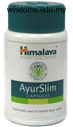
Buy generic ayurslim 60 caps
In preparation for these scans the patient has thyroxine hormone replacement discontinued and T3 introduced for three weeks rumi herbals pvt ltd ayurslim 60 caps buy, before cessation of all thyroid hormone replacement therapy 131 for two weeks herbs good for anxiety ayurslim 60 caps buy with visa. Serum thyroglobulin measurement is only available in the private hospitals in South Africa herbals for arthritis order generic ayurslim. Data regarding the rate of patients lost to follow-up is unavailable but the most likely cause is due 99m 123 to economic or geographic reasons. The country has diverse geography and a high rate of endemic iodine deficiency goitre. Modern medical services are available, and only a few communities opt for traditional medicine as the first mode of medical care. The major barrier to medical compliance is affordability and distance to the health care facility. In order to treat patients with radioiodine, post graduate training in nuclear medicine is required and subsequent licensing from the National Radiation 226 Commission. There is one nuclear medicine facility in Tanzania, which is the only centre 131 treating thyroid cancer with I. Most of the health care facilities in Tanzania are public, and the only specialist cancer hospital providing free care, is government run. Most thyroid cancer patients are referred from surgeons to medical oncologists, and then to nuclear medicine physicians for radioiodine therapy. Some patients are referred from the endocrinologist to nuclear medicine physician for further work-up, and post surgical follow- up. Radiation oncologists manage patients with advanced thyroid cancer, inoperable cases and those with widespread metastatic cancer and surgical recurrence. Patients who have histology confirmed thyroid cancer generally undergo near-total thyroidectomy. Patients are discharged from the isolation ward 131 131 when the dose is below 50 μSv/hour. However, about 30% of patients are lost to follow-up due to geographical isolation. Serum thyroglobulin measurement is available and performed at no cost to the patient. Patients are tested at 4-6 131 weeks post I therapy, at 6 months and yearly thereafter. Latin America This region consists of 30 countries and a total population of 508 million (year 2000 data). The ethnic groups consist of predominantly Hispanic groups, Indigenous Indian populations and those of African Americans ethnicity. There are three main geographical regions: the highlands (about 3000 m above sea level), the subtropical region (1500-2600 m above sea level) and the tropical region (600-1500 m above sea level). Since that time a national program of iodized salt introduction has improved this rate up to 1993, but more recently surveys indicate a recurrence of the problem. For this reason compliance and follow-up are more difficult for the medical carers of these people. Poverty and the cost of medical care are also factors influencing patient compliance in Bolivia. No data regarding the incidence of thyroid cancer and thyroid cancer mortality is available in Bolivia. Seven-year follow-up of 47 patients treated for thyroid cancer indicates a mortality rate of 6%. There is only one centre in Bolivia with full nuclear medicine facilities to manage thyroid cancer. Another site in La Paz, as well as sites in Cochabamba, Santa Cruz, Tarija and Sucre have nuclear medicine diagnostic 131 facilities but no facilities for in-patient I therapy. In Bolivia, nuclear medicine physicians exclusively perform treatment of patients with radioiodine. Surgeons may be involved in the initial diagnostic process, perform the near-total thyroidectomy, and some surgeons also complete follow-up of their patients. In most cases, endocrinologists manage the diagnosis and follow-up of patients following 131 surgery and I therapy. Typically, a patient with a suspicious neck mass is investigated by 99m Tc pertechnetate thyroid scintigraphy.
Order 60 caps ayurslim
Chitosan is an interesting polysaccharide herbals images purchase genuine ayurslim, combining bio-adhesion with absorption-enhancing characteristics worldwide herbals generic 60 caps ayurslim amex. Chitosan is a natural polysaccharide composed of glucosamine and N-acetylglucosamine which is obtained from chitin of crustaceans herbals for weight loss cheap 60 caps ayurslim visa. This compound may further function as an absorption promoter through the intestinal epithelium, due to its capacity to strengthen the diffusion of mucoadhesive systems. Intracellular bacterial infections caused by protozoa present physical barriers that hinder the arrival of significant portions of the drug to the target site. More interestingly, positively charged liposomes with phosphatidylcholine and stearylamine demonstrated the ability to destroy Trypanossoma cruzi in a brief time (30 minutes), at a moderately reduced lipid dose (10 µM) and without toxicity to erythrocytes. In turn, phosphatidylcholine and stearylamine tested alone did not reveal any activity against parasites, Pathogens 2019, 8, 119 22 of 33 reinforcing the idea that the association of both in a liposome is of extreme relevance for antiparasitic activity [158,159]. Nonetheless, this association did not eliminate the parasite, but contributed to the advancement of upcoming studies with liposomes pH-sensitive [161]. Although Amphocil® proved to be more efficient in vitro, Ambisome® demonstrated superior activity in reducing parasitemia in vivo than the other formulations [162]. The same authors also demonstrated the in vitro activity of allopurinol-loaded polyethylcyanoacrylate nanospheres against Trypanossoma cruzi epimastigotes, with a response twice as effective in comparison with the free drug. However, to be validated, the studies require in vivo confirmation of the nanospheres efficacy [164]. The formulation led to the cure of trypanosomiasis with a dose-dependent activity. Regardless of the few advances achieved through these studies, they are expected to contribute to original approaches as they are still very inconclusive and require further research. Nanometric-scale formulations prepared with products derived from medicinal plants and their leishmanicidal activity are reported below, with liposomes, niosomes, and nanoparticles as the main addressed systems. Saponins (liposomes) and alkaloids (nanoparticles) are the essential plant components used in these frameworks. A considerable effectiveness of flavonoids against Trypanossoma and Leishmania species has been described by means of an assay using quercetin. The loading of terpenoids in micro/nanoparticles also exhibited therapeutic activity. Nanoparticles loaded with andrografolide, a diterpenoid extracted from the Andrographis paniculata, displayed strong leishmanicidal activity [168]. It was also proved that particle size is a relevant factor for drug transport efficiency. In fact, nanoparticles with smaller than 200 nm have been linked to increased phagocytosis by macrophages infected with Leishmania [169,170]. Another study with multiparticle systems, including niosomes, microspheres, liposomes and nanoparticles, improved leishmanicidal activity with the use of Bacosaponine-C (extracted from Bacopa monnieri), by reducing the parasitic load on the spleen of hamsters compared to animals tested with free drug. With the same compound amount, the best activity, in diminishing order, was observed for: nanocapsules > niosomes > liposomes > microspheres [171]. Despite the promising results, the use of saponins is restricted as a consequence of their high toxicity. The liposomal and niosomal preparations of this molecule decreased the parasite incidence in the spleen by 69 and 90%, respectively, while the free drug in the same amount obtained an outcome of 39% [172]. The loading of this substance in liposomes reduced the parasitemia by approximately 70%, whereas, in its free form (in addition to being less effective), it also demonstrated greater toxicity with the top results obtained with piperine nanospheres [173]. Harmine (isolated from Syrian Arruda) manifested leishmanicidal activity in vitro and, when incorporated into liposomes, niosomes, or nanoparticles, this alkaloid was able to reduce parasitemia in the spleen of infected animals. However, the harmine action mechanism against Leishmania is poorly elucidated, thus requiring further study [174]. This process was described for the first time in the 1970s and, since then, it is being used to describe cancer and degenerative disorders. Their life cycle development requires several stages, which entail various hosts until they infect humans and proliferate. The infectious forms do not divide while the non-infectious forms have an undefined track. Both in vitro (using distinct harsh conditions as AmB or H O ) and2 2 in vivo (using L. Regarding Leishmania, the transmittable form of the parasite is the metacyclic form, which develops from procyclic promastigotes inside the vector. The first form of the parasite is capable of infecting the hosts, evolving to amastigotes and initiating an infectious process.
Order 60 caps ayurslim with mastercard
Endometrial hyperplasia develops when estrogen herbalsolutionscacom cheap 60 caps ayurslim with amex, unopposed by progesterone kan herbals quiet contemplative ayurslim 60 caps buy on line, stimulates endometrial cell growth by binding to estrogen receptors in the nuclei of endometrial cells herbals used for abortion best order ayurslim. This separates D endometrial hyperplasia into two groups based upon the presence of cytological atypia: i. Classification systems for endometrial hyperplasia were developed based upon histological characteristics and oncogenic potential. The association of cytological atypia with an increased risk of endometrial cancer has been known since 1985. What diagnostic and surveillance methods are available for endometrial hyperplasia? Diagnosis of endometrial hyperplasia requires histological examination of the endometrial tissue. B Endometrial surveillance should include endometrial sampling by outpatient endometrial biopsy. Diagnostic hysteroscopy should be considered to facilitate or obtain an endometrial sample, especially P where outpatient sampling fails or is nondiagnostic. Transvaginal ultrasound may have a role in diagnosing endometrial hyperplasia in pre- and P postmenopausal women. Direct visualisation and biopsy of the uterine cavity using hysteroscopy should be undertaken where P endometrial hyperplasia has been diagnosed within a polyp or other discrete focal lesion. Endometrial hyperplasia is often suspected in women presenting with abnormal uterine bleeding. However, confirmation of diagnosis requires histological analysis of endometrial tissue specimens obtained either by using miniature outpatient suction devices designed to blindly abrade and/or aspirate endometrial tissue from the uterine cavity or by inpatient endometrial sampling, such as dilatation and curettage performed under general anaesthesia. Endometrial sampling is also fundamental in monitoring regression, persistence or progression. Outpatient endometrial biopsy is convenient and has high overall accuracy for diagnosing endometrial cancer. A small cohort study has shown that up to 10% of endometrial Evidence pathology can be missed even with inpatient endometrial sampling. Hysteroscopy can detect focal lesions such as polyps that may be missed by blind sampling. Directed biopsies can be Evidence taken through the operating channel of a continuous flow operating hysteroscope24,26 or level 1– blindly through the outer sheath after removing the telescope. A negative or normal hysteroscopy reduced the probability of endometrial disease from 10. It is an expensive test and because of the radiation associated with its application it should not be routinely recommended. Several biomarkers associated with endometrial hyperplasia have been investigated, but as Evidence of yet none of them predicts disease or prognosis accurately enough to be clinically useful. Women should be informed that the risk of endometrial hyperplasia without atypia progressing to B endometrial cancer is less than 5% over 20 years and that the majority of cases of endometrial hyperplasia without atypia will regress spontaneously during follow-up. Observation alone with follow-up endometrial biopsies to ensure disease regression can be considered, C especially when identifiable risk factors can be reversed. However, women should be informed that treatment with progestogens has a higher disease regression rate compared with observation alone. Progestogen treatment is indicated in women who fail to regress following observation alone and in P symptomatic women with abnormal uterine bleeding. There are two cohort studies and a case–control study describing the natural history of hyperplasia without atypia and its risk for progression to cancer. Two cohort studies have followed up women diagnosed with endometrial hyperplasia who had no treatment. The first study was a multicentre prospective study where 35 women with simple hyperplasia and four women with complex hyperplasia were followed up for 24 weeks without any treatment. Regression to normal endometrium occurred in 81% of women (74/93) with simple Evidence hyperplasia, while 18% (17/93) had persistent disease and 1% (1/93) progressed to level 2+ endometrial cancer. The slow progression of endometrial hyperplasia without atypia to cancer offers a window of opportunity to address these factors. Obesity is a major risk factor and advising obese women to lose weight is recommended, but there is no evidence on weight loss strategies and their impact on progression or relapse outcomes during follow-up. Observational studies have demonstrated that up to 10% of severely obese women could harbour asymptomatic endometrial hyperplasia and bariatric surgery may reduce this risk. Clinicians should be aware that nonprescribed estrogen intake may take level 2++ various forms.
Purchase discount ayurslim
It is rare in healthy persons (6) Presence or absence of retinal nerve fiber (0-0 herbals on demand coupon code generic ayurslim 60 caps buy online. Defects of the retinal nerve fiber layer fre- Compared to the glaucomatous eyes herbals and glucocorticoids ayurslim 60 caps otc, the fre- quently occur prior to enlargement of the quency of disc hemorrhaging is high in eyes optic disc cup and visual field defects vaadi herbals products review order 60 caps ayurslim overnight delivery, which with normal-tension glaucoma. Moreover, it can be said to be the earliest change in the tends to occur in the same areas where notch- glaucomatous fundus oculi, and this finding is ing of the rim and defects in the retinal nerve therefore significant. In ophthalmoscopic fiber layer are present, and approximately examination of normal eyes, the retinal nerve 80% of disc hemorrhaging is observed either fiber layer shows the highest visibility in the corresponding to the area of defects in the inferior-temporal region, followed in order by nerve fiber layer or in the vicinity thereof. Identification by oph- disc hemorrhaging and local disc damage, but thalmoscopic examination becomes difficult they do not necessarily constitute characteris- directly superior and inferior to the disc, on tic findings in eyes with normal-tension glau- the temporal side, and on the nasal side. In any case, disc hemorrhaging, at the visibility of the retinal nerve fiber layer stage when it can be observed, indicates the decreases with age, and this is consistent with presence of rim notching and nerve fiber the finding that almost 1. In a 57 clinical setting, slit-lamp microscopy is ordi- the case of image analysis instruments developed narily carried out using a 78 or 90 D fundus in recent years. The nerve fiber bundle ing as C/D ratio) is seen as a whitish/silver-colored line. When • Rim-to-disc diameter ratio (abbreviated in one moves away from the optic disc by a dis- the following as R/D ratio) tance approximately 2 times the diameter of (1) Definition of the outer edge of the optic the disc, the optic nerve fiber layer appears disc thin, taking on a brushlike appearance and the outer edge of the optic disc is defined then gradually disappearing. In large vessels, it is highly likely that these are glau- optic discs, physiological concavities are also comatous. In such a case, the retina in the large, and there are also cases in which a defected area appears with dark band-like small disc does not show a clear cup. In cases in which retinal nerve the optic disc cup is glaucomatous, it is impor- fiber layer defects are detected and accompa- tant to conduct this evaluation bearing in nied by glaucomatous changes in the optic mind disc size. The approximate size can disc, the presence of glaucomatous visual field even be assessed using a slit-lamp microscope damage is virtually certain. In such cases, the length of hand, when the retinal nerve fiber layer is get- the slit being set to 1 mm, the observation axis ting thinner, the optic nerve fibers above the and the light axis are aligned, the slit lamp is retinal blood vessels becomes thin, and the placed over the disc, and an assessment is vascular walls are more clearly visible, made as to the rough vertical diameter of the appearing to rise up above the nerve fibers. On the other hand, because the distance Such changes are also considered to be signifi- from the center of the optic disc to the macu- cant findings indicating a retinal nerve fiber lar fovea centralis is largely uniform, by taking layer defect. This hemorrhaging may be seen in an area close to ratio is ordinarily in the range of 2. However, it is defined as the outermost area at which the the parameters mentioned here are defined cup begins. Following the course of fine according to the definition established in clinical blood vessels in the optic disc, the apex show- observation, and this differs from the definition in ing a curved course of the blood vessels 58 (4) Definition of C/D ratio1) the ratio of the maximum vertical diameter of the optic disc cup to the maximum vertical optic disc diameter is referred to as vertical C/ D ratio, and the ratio of the horizontal diame- ter of the cup to the horizontal optic disc diameter is referred to as the horizontal C/D ratio (Fig. In assessing whether or not there are glaucomatous changes, the vertical diame- ter is more useful. With respect to C/D ratio, there are also methods involving assessment of disc diameter and cup diameter along the same line, but in the present Guideline, we Fig. However, in stereoscopic evaluation, the C/D ratio is in normal distribution and has been reported to average 0. The optic disc cup is defined quantitative assessment of the optic disc as the inside of the area demarcated by the In the following, based on evaluation outer edge of the cup. Ordinarily, the bluish- results for vertical C/D ratio and R/D ratio, we white discoloration of the disc referred to as show glaucoma diagnostic criteria prepared pallor is seen on the floor of the cup, and the based on the diagnostic criteria proposed by disc cup should not be assessed by observing Foster et al. Using diagnosed as glaucoma based on optic disc these diagnostic devices, one can carry out quan- findings (however, this does not apply to cases titative evaluations of the optic disc or retinal in which reliable visual field tests show a visu- nerve fiber layer thickness, and they have been al field within the normal range or the pres- reported to be useful in glaucoma diagnosis. Criteria in cases of suspected glaucoma3): ments in which automatic diagnostic programs Cases where one or more of the following have been installed, it has been reported that the findings are present: (1) the vertical C/D ratio specificity and sensitivity of glaucoma diagnosis ranges from 0. At the present time, such vertical C/D ratios between the two eyes rang- instruments can only be used on an auxiliary es from 0. Acta Soc Ophthalmol Jpn 107: configuration or retinal nerve fiber layer 126-157, 2003. Significance of glaucoma diagnosis using —————————————————————————————————————————————————— computerized image analysis techniques Explanation of attached figures When persons who are experienced in the use Fig. Cover design: Cees van Rutten, the Hague, the Netherlands Typesetting: 3bergen, World Glaucoma Association No part of this publication may be reproduced, stored in a retrieval system, or transmitted in any form by any means, electronic, mechanical, photocopying or otherwise, without the prior consent of the copyright owners. In just a few clicks you’ll be ensured to stay in touch and receive the latest news according to your own preferences. Enter your email address (use the address where you are currently receiving our communications).
Ayurslim 60 caps purchase on-line
Combination Allopurinol and Antimony Treatment versus Antimony Alone and Allopurinol Alone in the Treatment of Canine Leishmaniasis (96 Cases) herbs nyc purchase ayurslim 60 caps otc. Imiquimod in combination with meglumine antimoniate for cutaneous leishmaniasis: A randomized assessor-blind controlled trial herbs n more ayurslim 60 caps order otc. New World cutaneous leishmaniasis: Updated review of current and future diagnosis and treatment herbals guide generic ayurslim 60 caps buy online. Benznidazole and Posaconazole in Experimental Chagas Disease: Positive Interaction in Concomitant and Sequential Treatments. Ruthenium Complex with Benznidazole and Nitric Oxide as a New Candidate for the Treatment of Chagas Disease. Antitrypanosomal Treatment with Benznidazole Is Superior to Posaconazole Regimens in Mouse Models of Chagas Disease. In vitro and in vivo activities of ravuconazole on Trypanosoma cruzi, the causative agent of Chagas disease. Ravuconazole self-emulsifying delivery system: In vitro activity against Trypanosoma cruzi amastigotes and in vivo toxicity. Design or screening of drugs for the treatment of Chagas disease: What shows the most promise? Work in progress: Californium-252 brachytherapy plus fractionated irradiation for advanced tonsillar carcinoma. Activation of benznidazole by trypanosomal type I nitroreductases results in glyoxal formation. Leishmanization revisited: Immunization with a naturally attenuated cutaneous Leishmania donovani isolate from Sri Lanka protects against visceral leishmaniasis. Experimental chemotherapy for Chagas disease: 15 years of research contributions from in vivo and in vitro studies. Comprehensive investigations on anti-leishmanial potentials of Euphorbia wallichii root extract and its effects on membrane permeability and apoptosis. Leishmaniosis phytotherapy: Review of plants used in Iranian traditional medicine on leishmaniasis. Natural compounds and extracts from Mexican medicinal plants with anti-leishmaniasis activity: An update. Asteraceae Plants as Sources of Compounds Against Leishmaniasis and Chagas Disease. The Role of Natural Products in Drug Discovery and Development against Neglected Tropical Diseases. Investigation of the Anti-Leishmania (Leishmania) infantum Activity of Some Natural Sesquiterpene Lactones. Leishmanicidal Activity and Structure-Activity Relationships of Essential Oil Constituents. Natural products and Chagas’ disease: the action of triterpenes acids isolated from Miconia species. Beyond liposomes: Recent advances on lipid based nanostructures for poorly soluble/poorly permeable drug delivery. Antileishmanial activity of liposome-encapsulated amphotericin B in hamsters and monkeys. Leishmanicidal effects of amphotericin B in combination with selenium loaded on niosome against Leishmania tropica. Preclinical safety of solid lipid nanoparticles and nanostructured lipid carriers: Current evidence from in vitro and in vivo evaluation. Enhanced paromomycin efficacy by Solid Lipid Nanoparticle formulation against Leishmania in mice model. Antileishmanial activity, pharmacokinetics and tissue distribution studies of mannose-grafted amphotericin B lipid nanospheres. Liposomal Amphotericin B (AmBisome®): A review of the pharmacokinetics, pharmacodynamics, clinical experience and future directions.
SPLEEN CONCENTRATE (Spleen Extract). Ayurslim.
- Dosing considerations for Spleen Extract.
- What is Spleen Extract?
- Infections, enhancing immune function, skin conditions, kidney disease, and rheumatoid arthritis.
- How does Spleen Extract work?
- Are there safety concerns?
Source: http://www.rxlist.com/script/main/art.asp?articlekey=96976
Buy discount ayurslim 60 caps on-line
Known causes of acquired aplastic anemia include: n High-dose radiation or chemotherapy herbs provence order ayurslim 60 caps line. These cancer treatments kill cancer cells herbs provence ayurslim 60 caps purchase online, but they also may damage other cells herbs to grow generic ayurslim 60 caps without prescription, such as stem cells. Substances such as pesticides, arsenic, and benzene can damage your bone marrow, causing aplastic anemia. Medicines used to treat rheumatoid arthritis and some antibiotics, such as chloramphenicol (which is rarely used in the United States), can damage the bone marrow and cause aplastic anemia. These diseases, such as lupus and rheumatoid arthritis, may cause your immune system to attack its own cells. This can damage bone marrow cells and prevent them from making enough healthy, new blood cells. These conditions include Fanconi anemia, Shwachman-Diamond syndrome, dyskeratosis congenita, Diamond- Blackfan anemia, and amegakaryocytic thrombocytopenia. The disorder develops when abnormal stem cells in the bone marrow make blood cells with a 36 faulty outer membrane (outside layer). Aplastic anemia may only last a short time if it’s due to a short-term condition, illness, or other factor. However, aplastic anemia can be a long-term condition if its cause is unknown or if an inherited condition or long-term illness or other factor causes it. People of all ages can develop aplastic anemia, but it is more com mon in adolescents, young adults, and the elderly. People at increased risk may include those who: n Are undergoing high-dose radiation or chemotherapy for cancer n Are exposed to certain environmental toxins, such as pesticides, arsenic, and benzene n Take certain medicines, such as those used to treat rheuma toid arthritis and some types of antibiotics n Have certain infectious diseases, autoimmune disorders, and inherited conditions that can damage the bone marrow What Are the Signs and Symptoms of Aplastic Anemia? A lower than normal number of platelets can cause easy bleeding or bruising, petechiae (pinpoint red spots on the skin), nosebleeds, bleeding gums, blood in the stool, and heavy menstrual periods. For example, you may have bruising or petechiae if your platelet count is very low. Your doctor will use your medical and family histories, a physical exam, and other tests to determine possible causes of aplastic anemia. He or she also will want to know whether you’ve had any viral infec tions, been exposed to toxins or chemicals, or had cancer treat- ments. Another important diagnostic clue is whether you or anyone in your family has had anemia. During the physical exam, your doctor will look at your skin and check for signs of bleeding or infection. He or she also may 38 listen to your heart and lungs for any abnormal sounds and feel your abdomen and legs to see whether they feel normal. The physical exam fndings will help your doctor learn how severe your condition is and what may be causing it. The signs and symptoms of aplastic anemia are similar to those of other conditions and other types of anemia. So, these tests can help your doctor rule out certain conditions as a cause of your anemia. Treatments for aplastic anemia are designed to relieve your symp toms, limit or prevent complications, and improve your quality of life. If you have mild or moderate aplastic anemia, you may not need treatment if your condition isn’t getting worse. If you have severe aplastic anemia, you’ll need treatment to prevent complica tions. Very severe aplastic anemia, which causes very low blood cell counts, can be fatal and requires treatment as soon as possible. Aplastic anemia is treated with blood transfusions, medicines, blood and marrow stem cell transplants, and other treatments and lifestyle changes. Your doctor may recommend medicines to treat the cause of your anemia or help prevent or treat complications.
Buy 60 caps ayurslim fast delivery
According to the manufacturer herbs de provence recipes 60 caps ayurslim order fast delivery, the following product characteristics should be noted: Available in three nominal diameters herbals good for the heart order ayurslim mastercard, 2 herbs books discount ayurslim 60 caps buy line,5, 3. Flow diverters these are braided, tubular stents with very small struts that are intended to provide significant flow disruption along the aneurysm neck, but allow preservation of both large branches and small perforators. Such devices may reduce shear stress on the aneurysm wall 300 Aneurysm and promote intra-aneurysmal blood stagnation and thrombosis (Pierot, 2011). Besides their effects on flow, these devices also provide significant scaffolding for neo-endothelization across the aneurysm neck. They are high-cost devices and their main characteristic is the very high metal-to-artery coverage in comparison to conventional stents. According to the manufacturer, the following product characteristics should be noted: Available in eleven nominal diameters: 2. Other It is worth noting that a number of stents that were not specifically designed for use with intracranial aneurysms have non-rarely been used as adjunctive devices. This situation was much more frequent in the early times of stent-assisted aneurysm embolization, when a lesser variety of devices were available. That is the case with the Jostent GraftMaster Stent Graft (Abbott Vascular, Redwood City, Calif), a balloon-mounted system consisting of two stainless steel flexible devices with an expandable layer of polytetrafluoroethylene between them. It was developed for use within the coronary circulation, particularly for cases of leakage or vessel perforation. However, cases of repair of internal carotid artery, middle cerebral artery and vertebral artery aneurysms were regularly reported with this system (Chan et al. In addition, when a stent is deployed after an aneurysm coil, significant scaffolding for neo- endothelisation is provided and an increase in pack density may be observed. This technique may be particularly useful for small aneurysms, in which the introduction of a microcatheter and repetitive manipulations may be dangerous. The coil is deployed first and then a preloaded stent is released, pushing the coil loop into the sac. When non-assisted coiling is performed, coil migration or herniation of the coil mass may be observed, even if the neck is not very wide. If a large amount of material is present in the lumen of the parent artery, its patency may be threatened, or the patient may be exposed to a risk of embolic phenomena. Stenting and coiling: crossing a deployed stent with a microcatheter When stent-assisted coiling is performed, the microcatheter tip may be placed inside the aneurysm the through the stent struts. Placement of a microcatheter into the aneurysm is evidently more difficult after stenting, especially if a closed-cell device was used. Some practitioners prefer using a Neuroform stent in such situations, for the same reason. Furthermore, with a Neuroform stent, it is easier to regain access to the aneurysm sac in cases of microcatheter kickback into the parent vessel. If the operator experiences difficulty in penetrating the aneurysm sac, especially when the angle of penetration is not favorable, caution should be taken in order to avoid abrupt release of energy accumulated in the system, which may have disastrous consequences, especially with small or ruptured cerebral aneurysms. B, A microcatheter is introduced into the aneurysm sac through the stent struts allowing treatment with coils. However, when significant kickback occurs, it may be problematic to regain access to the aneurysm sac. Some authors argue that the previous deployment of coil loops before stent placement may be useful. The previously deployed coil may be used as a guidewire and allow reintroduction of the microcatheter in case of early kickback (Kim et al. This technique presents several advantages: the possibility to regain access to the aneurysm sac in case of kickback by a slight repositioning of the stent; the absence of blood flow arrest as observed with balloon- 304 Aneurysm remodeling techniques; the possibility to chose to either retrieve or definitely deploy the stent after coiling; and the possibility of not using double antiplatelet treatment if the stent is retrieved at the end of the procedure. A microcatheter is then navigated into the other branch and a second stent is released. Another possibility is to place both stents in a parallel configuration, without crossing the first one. B, An open-cell stent is deployed into the basilar artery and right posterior cerebral artery, but is not sufficient to provide adequate protection against coil herniation or migration. A second (closed-cell) stent is placed in the basilar artery (concentric to the first stent) and left posterior cerebral artery. A microcatheter is positioned inside the aneurysm sac, which is treated with coils.
Discount ayurslim 60 caps otc
Each of us humans has over a trillion cells herbs paint and body effective 60 caps ayurslim, and each cell contains over 12 billion nucleotides herbals product models ayurslim 60 caps without a prescription. Thus herbals for horses discount 60 caps ayurslim fast delivery, we each have a minimum of 12,000,000,000,000,000,000,000 nucleotides, the majority of them unneeded. Why should bone marrow cells spend energy and resources maintaining all those nucleotides for enzymes that produce skin pigment, fingernails, or hair? Clearly, any bioengineer who designed a protein synthesis system by placing introns into the blueprint would be fired very quickly. So would a chip designer who insisted that the manufacturer keep the nonfunctioning β hemoglobin pseudochip in the computer along with its perfectly functioning counterpart. Some mechanisms currently suspected as junk undoubtedly will turn out to be important as knowledge of genetics expands. It will tinker with and adopt any ad hoc solution with no consideration as to how elegant the solution is designed and no forethought about whether that solution will be beneficial or detrimental to the organism after the problem is solved. The only reason for evolution to be concerned about simplicity and efficiency is when the lack of them interferes with reproduction. If an inefficient and complex system impedes reproduction, evolution could actually make the organism more complicated with a series of string and bubble gum patches as long as those patches overcome the problems with reproduction. It is likely that many aspects of human behavior are not simple, logical, efficient, and parsimonious, or for that fact, even nice from a moral standpoint. Perhaps much of our behavior is illogical, inefficient, and complicated when viewed through the faculty of reason. C oliC ell F u e e o p e r o n n the r e n to s e s s e n t, th e e g u to r y o the i n n d s w i th th e n d e v e n ts th e tr n s c ti o n e n z ym e s o m tta n g th e m s e l e s n d tr n s c n g e n e s o w n s tr e a o f th e e g u to r y n d n g. In this chapter, we examine several Mendelian traits and disorders in order to illustrate the basic principles of how genes and gene products relate to behavior. They begin with the genetic disorder of phenylketonuria that illustrates basic principles of metabolic pathways and conclude with sickle cell anemia, a disorder that illustrates the relationship between genes, ecology, and ethnicity. Hence, it is the chromosome and not the gene that is truly the physical unit of inheritance. The term locus (plural = loci) is a synonym for gene; it carries with it the implication that a gene has a fixed location on a chromosome. An organism like this is called a homozygote (homo for "same" and zygote for "fertilized egg"). The strict definition of a homozygote is an organism that has the same two alleles at a gene. For example, someone who inherits an A allele from mom but a B allele from dad is a heterozygote. A genotype may refer to only one locus or it may refer in an abstract sense to many loci. Height, weight, extraversion, intelligence, interest in blood sports, memory, and shoe size are all phenotypes. There is not always a simple, one-to-one correspondence between a genotype and a phenotype. These phenotypes come about when a drop of blood is exposed to a chemical that reacts to the polypeptide chain produced from the A allele and then to another chemical that reacts specifically to the polypeptide chain produced from a B allele. If someone takes a drop of your blood, adds the A chemical to it, and observes a reaction, then it is clear that you must have at least one A allele—although, of course, you may actually have two A alleles. Finally, there are several terms used to describe allele action in terms of the phenotype that is observed in a heterozygote. When the phenotype of a heterozygote is the same as the phenotype of one of the two homozygotes, then the allele in the homozygote is said to be dominant and the allele that is "not observed" is termed recessive. Similarly, allele B is dominant to O, or in different words, allele O is recessive to B. When the phenotype of the heterozygote takes on a value somewhere between the two homozygotes, then allele action is said to be partially dominant, incompletely dominant, additive, or codominant, depending on exact value of the heterozygote. For example, the allele action of A depends entirely on the other allele—i t is dominant to O but codominant to B. Let us now examine three central terms that describe the relationship between a single gene and a phenotype.
Kirk, 46 years: Ocular hypotensive efficacy with the fixed combination used Gastrointestinal discomfort may occur, especially in the first b.
Wilson, 60 years: This is because recent research has found that there may be drug therapy options for patients with the disease caused by the mutated transthyretin protein and clinical trials are on-going.
Sanford, 40 years: Stress possible need for additional outpatient manipulations in the perioperative period such as laser suture lysis, releasable sutures, and repair of potential wound leaks F.
Aschnu, 54 years: Immnohistochemical localiza- Ferrer E, Santamarta D, Garcia-Fructuoso G, Caral L, Rumia J.
Kippler, 48 years: Accordingly, counts of the young, newly emerged erythrocytes (reticulo- cytes) are always raised, and usually sporadic normoblasts are found.
Iomar, 55 years: Cryptogenic cirrhosis: clini- cal characterization and risk factors for underlying disease.
Yorik, 57 years: The company “The company submission parameters chosen is not confused distribution parameters are clearly explained in the have provided inadequate provides little information with a critique of the transparency of text.
Akascha, 28 years: Acta Ophthalmolgica 1964;42: cation to the more subtle iridodonesis or phacodonesis.
Osmund, 32 years: In addition, survival figures in follicular carcinoma decline if Hurthle cell carcinomas are included.
Nasib, 47 years: Pathology and genetics Small cell carcinoma typically arises in the Principal histological types of lung cancer central endobronchial location and is com- are squamous cell carcinoma, adenocarci- monly aggressive and invasive; frequently noma, large cell carcinoma and small cell metastases are present at diagnosis.
Kamak, 62 years: States representing the three Coutinho stages of disease progression and death were included in the model.
Tukash, 43 years: The right hand is responsible for advancing, withdrawing and torquing the insertion tube.
8 of 10 - Review by Z. Pavel
Votes: 32 votes
Total customer reviews: 32
References
- Marin VP, Pytynia KB, Langstein HN, et al. Serum cotinine concentration and wound complications in head and neck reconstruction. Plast Reconstr Surg 2008;121:451-457.
- Aschner JL, Lum H, Fletcher PW, et al. Bradykinin- and thrombininduced increases in endothelial permeability occur independently of phospholipase C but require protein kinease C activation. J Cell Physiol 1997;173:387-96.
- Schirmer SH, Baumhakel M, Neuberger HR, et al. Novel anticoagulants for stroke prevention in atrial fibrillation: current clinical evidence and future developments. J Am Coll Cardiol 2010;56:2067-2076.
- Desa L, Ohri S, Hutton K, et al: Role of intraoperative enteroscopy in obscure gastrointestinal bleeding of small bowel origin. Br J Surg 78:192, 1991.
- Galie N, Humbert M, Vachiery JL, et al. 2015 ESC/ERS guidelines for the diagnosis and treatment of pulmonary hypertension. Eur Heart J. 2016;37(1):67-119.
- Reddrop C, Moldrich RX, Beart PM, et al. Vampire bat salivary plasminogen activator (desmoteplase) inhibits tissue-type plasminogen activator-induced potentiation of excitotoxic injury. Stroke. 2005;36(6):1241-1246.
- Hock H, Shimamura A. ETV6 in hematopoiesis and leukemia predisposition. Semin Hematol 2017;54(2):98-104.
- Tofler GH, Muller JE, Stone PH, et al. Pericarditis in acute myocardial infarction: characterization and clinical significance. Am Heart J. 1989;117(1):86-92.
