Peter J McDonnell, M.D.
- Director of The Wilmer Eye Institute
- Professor of Ophthalmology

https://www.hopkinsmedicine.org/profiles/results/directory/profile/0003333/peter-mcdonnell
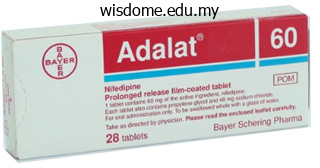
Adalat dosages: 30 mg, 20 mg
Adalat packs: 30 pills, 60 pills, 90 pills, 120 pills, 180 pills, 270 pills, 360 pills
Discount adalat 20 mg buy line
Thus heart attack quick treatment buy adalat paypal, by flowing slowly to preserve the countercurrent mechanism arteria facial adalat 30 mg purchase with amex, as the vasa recta descend arteria haemorrhoidalis media order adalat australia, Na and urea are + freely able to enter the capillary, while water freely leaves; as they ascend, Na and urea are secreted into the surrounding medulla, while water reenters and is removed. The movement of Na out of the lumen – of the collecting duct creates a negative charge that promotes the movement of Cl out of the lumen into the interstitial space by a paracellular route across tight junctions. Peritubular capillaries receive the solutes and water, returning them to the circulation. In addition, as Na is pumped out of the cell, the resulting electrochemical gradient attracts ++ Ca into the cell. Finally, calcitriol (1,25 dihydroxyvitamin D, the active form of vitamin D) is very important for calcium ++ recovery. It induces the production of calcium-binding proteins that transport Ca into the cell. These binding proteins are also important for the movement of calcium inside the cell and aid in exocytosis of calcium across the basolateral ++ membrane. Collecting Ducts and Recovery of Water Solutes move across the membranes of the collecting ducts, which contain two distinct cell types, principal cells and intercalated cells. A principal cell possesses channels for the recovery or loss of sodium and potassium. As in other portions of the nephron, there is an array of micromachines (pumps and channels) on display in the membranes of these cells. Regulation of urine volume and osmolarity are major functions of the collecting ducts. By varying the amount of water that is recovered, the collecting ducts play a major role in maintaining the body’s normal osmolarity. If the blood becomes hyperosmotic, the collecting ducts recover more water to dilute the blood; if the blood becomes hyposmotic, the collecting ducts recover less of the water, leading to concentration of the blood. Another way of saying this is: If plasma osmolarity rises, more water is recovered and urine volume decreases; if plasma osmolarity decreases, less water is recovered and urine volume increases. As the ducts descend through the medulla, the osmolarity surrounding them increases (due to the countercurrent mechanisms described above). If aquaporin water channels are present, water will be osmotically pulled from the collecting duct into the surrounding interstitial space and into the peritubular capillaries. By altering the number of aquaporin channels, the volume of water recovered or lost is altered. This, in turn, regulates the blood osmolarity, blood pressure, and osmolarity of the urine. By also stimulating aldosterone production, it provides a longer-lasting mechanism to support blood pressure by maintaining vascular volume (water recovery). As + + + the pump recovers Na for the body, it is also pumping K into the forming urine, since the pump moves K in the opposite + + direction. When aldosterone decreases, more Na remains in the forming urine and more K is recovered in the circulation. Still other channels in the principal cells secrete K into the collecting duct + in direct proportion to the recovery of Na. This function lowers the acidity of the plasma while increasing the acidity of the urine. This rate determines how much solute is retained or discarded, how much water is retained or discarded, and ultimately, the osmolarity of blood and the blood pressure of the body. Sympathetic Nerves the kidneys are innervated by the sympathetic neurons of the autonomic nervous system via the celiac plexus and splanchnic nerves. Reduction of sympathetic stimulation results in vasodilation and increased blood flow through the kidneys during resting conditions. When the frequency of action potentials increases, the arteriolar smooth muscle constricts (vasoconstriction), resulting in diminished glomerular flow, so less filtration occurs. Under conditions of stress, sympathetic nervous activity increases, resulting in the direct vasoconstriction of afferent arterioles (norepinephrine effect) as well as stimulation of the adrenal medulla. The adrenal medulla, in turn, produces a generalized vasoconstriction through the release of epinephrine. This includes vasoconstriction of the afferent arterioles, further reducing the volume of blood flowing through the kidneys.
Adalat 30 mg order visa
However blood pressure medication causing low blood pressure cheap adalat 20 mg without prescription, in addition to low oxygen pulse pressure 28 order adalat 30 mg, high carbon dioxide heart attack follow me quality 20 mg adalat, and low pH levels, there appears to be a complex interplay of factors related to the nervous system and the respiratory centers of the brain. First, a conscious decision to partake in exercise, or another form of physical exertion, results in a psychological stimulus that may trigger the respiratory centers of the brain to increase ventilation. In addition, the respiratory centers of the brain may be stimulated through the activation of motor neurons that innervate muscle groups that are involved in the physical activity. Finally, physical exertion stimulates proprioceptors, which are receptors located within the muscles, joints, and tendons, which sense movement and stretching; proprioceptors thus create a stimulus that may also trigger the respiratory centers of the brain. These neural factors are consistent with the sudden increase in ventilation that is observed immediately as exercise begins. Because the respiratory centers are stimulated by psychological, motor neuron, and proprioceptor inputs throughout exercise, the fact that there is also a sudden decrease in ventilation immediately after the exercise ends when these neural stimuli cease, further supports the idea that they are involved in triggering the changes of ventilation. High Altitude Effects An increase in altitude results in a decrease in atmospheric pressure. Although the proportion of oxygen relative to gases in the atmosphere remains at 21 percent, its partial pressure decreases (Table 22. As a result, it is more difficult for a body to achieve the same level of oxygen saturation at high altitude than at low altitude, due to lower atmospheric pressure. In fact, hemoglobin saturation is lower at high altitudes compared to hemoglobin saturation at sea level. For example, hemoglobin saturation is about 67 percent at 19,000 feet above sea level, whereas it reaches about 98 percent at sea level. Partial Pressure of Oxygen at Different Altitudes Example Altitude (feet above Atmospheric Partial pressure of location sea level) pressure (mm Hg) oxygen (mm Hg) New York City, New 0 760 159 York Boulder, Colorado 5000 632 133 Aspen, Colorado 8000 565 118 Table 22. A lower partial pressure of oxygen means that there is a smaller difference in partial pressures between the alveoli and the blood, so less oxygen crosses the respiratory membrane. Despite this, the tissues of the body still receive a sufficient amount of oxygen during rest at high altitudes. First, the number of oxygen molecules that enter the tissue from the blood is nearly equal between sea level and high altitudes. At sea level, hemoglobin saturation is higher, but only a quarter of the oxygen molecules are actually released into the tissue. At high altitudes, a greater proportion of molecules of oxygen are released into the tissues. Physical exertion, such as skiing or hiking, can lead to altitude sickness due to the low amount of oxygen reserves in the blood at high altitudes. At sea level, there is a large amount of oxygen reserve in venous blood (even though venous blood is thought of as “deoxygenated”) from which the muscles can draw during physical exertion. Because the oxygen saturation is much lower at higher altitudes, this venous reserve is small, resulting in pathological symptoms of low blood oxygen levels. You may have heard that it is important to drink more water when traveling at higher altitudes than you are accustomed to . This is because your body will increase micturition (urination) at high altitudes to counteract the effects of lower oxygen levels. By removing fluids, blood plasma levels drop but not the total number of erythrocytes. In this way, the overall concentration of erythrocytes in the blood increases, which helps tissues obtain the oxygen they need. Acclimatization is the process of adjustment that the respiratory system makes due to chronic exposure to a high altitude. Over a period of time, the body adjusts to accommodate the lower partial pressure of oxygen. The low partial pressure of oxygen at high altitudes results in a lower oxygen saturation level of hemoglobin in the blood. With more red blood cells, there is more hemoglobin to help transport the available oxygen. Even though there is low saturation of each hemoglobin molecule, there will be more hemoglobin present, and therefore more oxygen in the blood. It is a complex process that includes many structures, most of which arise from the endoderm.
Cheap adalat 30 mg mastercard
It is not by accident that several of the new behavior therapies are most closely linked to this wing of behavior therapy hypertension foods 20 mg adalat buy overnight delivery, which only recently has developed sufficiently to impact adult psychotherapy in a powerful way arterial insufficiency buy adalat paypal. Behavior analysis is much easier to understand when its philosophical foundations are understood arrhythmia qt prolongation buy generic adalat 20 mg line. Although mechanistic forms of behavior analysis exist, by far the more dominant strand of modern behavior analysis is based on a type of American pragmatism we have termed functional contextualism (Hayes, 1993). A full discussion of contextualism as a philosophy of science is a topic beyond the scope of the present chapter (but see Biglan & Hayes, 1996; Hayes et al. Second, this philosophical difference seems to make more sense of the difference between secondgeneration behavior therapy and the new forms that have emerged. Acceptance and Commitment Therapy 7 the contextualistic wing of the new behavior therapies conceptualizes psychological events as a set of ongoing interactions between whole organisms and historically and situationally defined contexts. The root metaphor of contextualism (Pepper, 1942) is the “ongoing act in context,” that is, the commonsense situated action. Contextualists seek to maintain contact with the whole event and its context, and to analyze that event in such a way that its holistic quality is not undermined. Contextualists are supremely interested in function over form, because formal events literally have no meaning. An event disconnected from its history and current situational context is, in some sense, not an “event” at all: “It is not an act conceived as alone or cut off that we mean; it is an act in and with its setting” (Pepper, 1942, p. Mechanists deal with functional events by assembling a composite from the “elementary” pieces of interest. The assumptions of mechanism lead to the idea that the world is preorganized into parts, relations, and forces—one only has to discover the true underlying elements. Thus, an ontological claim underlies mechanism: the parts are already there; we must find them; without them, we cannot understand complexity. A functional unit is the unit, but it is so for pragmatic purposes brought into the situation by the analyst. Just as “going to the store” can be a functional unit, so too can “analyzing patients behavior into treatment responsive units. From the point of view of contextualism, determining the functional nature of a given event requires an ever-widening examination of context. The movement of a leg that occurs in the context of particular sequences of leg movements is “walking,” whereas the same movement in another context is “kicking. Making a dinner in the context of having one’s boss visit is different than making a private dinner to be eaten alone. What limits the process of examining context in an ever widening circle is the contextualist’s pragmatic view of truth. The process of contextual explication is not thought of as “discovering” the “truth” but as a process of construing the situation so that effective action is possible. Thus, analysis for a contextualist “becomes important in reference to the end” (Pepper, 1942, p. Skinner is quite clear about this: “It is true that we could trace human behavior not only to the physical conditions which shape and maintain it but also to the causes of those conditions and the causes of those causes, almost ad infinitum” but we need take analysis only to the point at which “effective action can be taken” (Skinner, 1974, p. Thus, a “proposition is ‘true’ to the extent that with its help the listener responds effectively to the situation it describes” (p. That stance on truth, built into behavior analysis, has a big impact on treatments that take a functional analytic approach. It is also this pragmatic approach that makes goals so important in contextualism. In order to know whether one is responding effectively, it is necessary to know what effects are being sought. Thus, goals are foundational in contextualism, and different goals can lead to different types of contextualism (Hayes, 1993). Goals enable analysis by allowing successful working to be assessed, but goals can only be stated, not evaluated.
Adalat 20 mg buy without prescription
In actuality arteria japan cheap adalat 30 mg without prescription, there are some elements of the peripheral nervous system that are within the cranial or vertebral cavities arteria facialis buy adalat 20 mg fast delivery. The peripheral nervous system is so named because it is on the periphery—meaning beyond the brain and spinal cord ulterior motive synonym discount adalat uk. Depending on different aspects of the nervous system, the dividing line between central and peripheral is not necessarily universal. A glial cell is one of a variety of cells that provide a framework of tissue that supports the neurons and their activities. The neuron is the more functionally important of the two, in terms of the communicative function of the nervous system. To describe the functional divisions of the nervous system, it is important to understand the structure of a neuron. Neurons are cells and therefore have a soma, or cell body, but they also have extensions of the cell; each extension is generally referred to as a process. There is one important process that every neuron has called an axon, which is the fiber that connects a neuron with its target. Looking at nervous tissue, there are regions that predominantly contain cell bodies and regions that are largely composed of just axons. These two regions within nervous system structures are often referred to as gray matter (the regions with many cell bodies and dendrites) or white matter (the regions with many axons). The colors ascribed to these regions are what would be seen in “fresh,” or unstained, nervous tissue. It can be pinkish because of blood content, or even slightly tan, depending on how long the tissue has been preserved. But white matter is white because axons are insulated by a lipid-rich substance called myelin. Lipids can appear as white (“fatty”) material, much like the fat on a raw piece of chicken or beef. Actually, gray matter may have that color ascribed to it because next to the white matter, it is just darker—hence, gray. The distinction between gray matter and white matter is most often applied to central nervous tissue, which has large regions that can be seen with the unaided eye. When looking at peripheral structures, often a microscope is used and the tissue is stained with artificial colors. There is also a potentially confusing use of the word ganglion (plural = ganglia) that has a historical explanation. In the central nervous system, there is a group of nuclei that are connected together and were once called the basal ganglia before “ganglion” became accepted as a description for a peripheral structure. Some sources refer to this group of nuclei as the “basal nuclei” to avoid confusion. There is an important point to make about these terms, which is that they can both be used to refer to the same bundle of axons. The most obvious example of this is the axons that project from the retina into the brain. Those axons are called the optic nerve as they leave the eye, but when they are inside the cranium, they are referred to as the optic tract. There is a specific place where the name changes, which is the optic chiasm, but they are still the same axons (Figure 12. The same axons extend from the eye to the brain through these two bundles of fibers, but the chiasm represents the border between peripheral and central. This is a tool to see the structures of the body (not just the nervous system) that depends on magnetic fields associated with certain atomic nuclei. The utility of this technique in the nervous system is that fat tissue and water appear as different shades between black and white. How do the imaging techniques shown in this game indicate the separation of white and gray matter compared with the freshly dissected tissue shown earlier?
Cheap adalat 20 mg on-line
The epidermis is attached to the inner arrhythmia nclex purchase adalat canada, thicker blood pressure 50 over 30 cheap adalat 20 mg without a prescription, connective tissue part called the dermis heart attack move me stranger extended version adalat 20 mg purchase mastercard. This layer, also called the superficial fascia or hypodermis, consists of areolar and adipose tissues. Fibbers from the dermis extend down into the subcutaneous layer and anchor the skin to it. In response to low environmental temperature, production of sweat is decreased, which helps conserve heat. These are made available to the body when there is depletion which may be due to starvation. In moderate exercise, skin blood flow may increase, which helps dissipate heat from the body. During hard exercise, however, skin blood vessels constrict (narrow) somewhat, and more blood is able to circulate to contracting muscles. Enzymes in the liver and kidneys then modify the molecule, finally producing calcitriol; the most active form of vitamin D. Calcitriol contributes to the homeostasis of body fluids by aiding absorption of calcium in foods. According to the synthesis sequence just described, vitamin D is a hormone, since it is produced in one location in the body, transported by the blood, and then exerts its effect in another location. They produce the protein keratin that helps waterproof and protect the skin and underlying tissues. Their long, slender projections extend between and transfer granules of melanin to keratinocytes. These cells are located in the deepest layer (stratum basale) of the epidermis of hairless skin, where they are attached to keratinocytes by desmosomes. Merkel cells make contact with the flattened portion of the ending of a sensory neuron (nerve cell), called a tactile (Merkel) disc, and are thought to function in the sensation of touch. Where exposure to friction is greatest, such as in the palms and soles, the epidermis is thicker (l to 2 mm) and has five layers. Constant exposure of thin or thick skin to friction or pressure stimulates formation of a callus, an abnormal thickening of the epidermis. Stratum basale: This single layer of cuboidal to columnar cells contains stem cells, which are capable of continued cell division, and Melanocytes. The stratum basale also contains tactile (Merkel) discs that are sensitive to touch. The cells here appear to be covered with prickly spines (spinosum prickly) because the cells shrink apart when the tissue is prepared for microscopic examination. This compound is the precursor of keratin, a protein found in the outer layer of the epidermis. Keratin forms a barrier that protects deeper layers from injury and microbial invasion and makes the skin waterproof. It consists of three to five rows of clear, flat, dead cells that contain droplets of an intermediate substance that is formed from keratohyalin and is eventually transformed to keratin. This layer consists of 25 to 30 rows of flat, dead cells completely filled with keratin. The stratum corneum serves as an effective barrier against light and heat waves, bacteria, and many chemicals. In the process of keratinization, cells newly formed in the basal layers undergo a developmental process as they are pushed to the surface. At the same time the cytoplasm, nucleus, and other organelles disappear, and the cells die. Eventually, the keratinised cells slough off and are replaced by underlying cells that, in tum, become keratinised. The whole process by which a cell forms in the basal layer, rises to the surface, becomes keratinised, and sloughs off takes two to four weeks. The dermis is very thick in the palms and soles and very thin in the eyelids, penis, and scrotum. It also tends to be thicker on the dorsal than the ventral aspects of the body and thicker on the lateral than the medial aspects of the extremities.
Levant Nut (Levant Berry). Adalat.
- How does Levant Berry work?
- Abnormal movements of the eyeball, dizziness, scabies, lice, epilepsy, night sweats, use as a stimulant, and malaria.
- Dosing considerations for Levant Berry.
- Are there safety concerns?
- What is Levant Berry?
Source: http://www.rxlist.com/script/main/art.asp?articlekey=96548
Adalat 20 mg buy overnight delivery
There are three dilations in the wall of the aorta just superior to the aortic semilunar valve heart attack questions discount adalat american express. Two of these zero pulse pressure generic 30 mg adalat free shipping, the left posterior aortic sinus and anterior aortic sinus blood pressure 210 over 110 adalat 20 mg purchase mastercard, give rise to the left and right coronary arteries, respectively. The third sinus, the right posterior aortic sinus, typically does not give rise to a vessel. Coronary vessel branches that remain on the surface of the artery and follow the sulci are called epicardial coronary arteries. The left coronary artery distributes blood to the left side of the heart, the left atrium and ventricle, and the interventricular septum. The circumflex artery arises from the left coronary artery and follows the coronary sulcus to the left. Along the way it gives rise to numerous smaller branches that interconnect with the branches of the posterior interventricular artery, forming anastomoses. An anastomosis is an area where vessels unite to form interconnections that normally allow blood to circulate to a region even if there may be partial blockage in another branch. Therefore, this ability is somewhat restricted in the heart so a coronary artery blockage often results in death of the cells (myocardial infarction) supplied by the particular vessel. The right coronary artery proceeds along the coronary sulcus and distributes blood to the right atrium, portions of both ventricles, and the heart conduction system. The marginal arteries supply blood to the superficial portions of the right ventricle. On the posterior surface of the heart, the right coronary artery gives rise to the posterior interventricular artery, also known as the posterior descending artery. It runs along the posterior portion of the interventricular sulcus toward the apex of the heart, giving rise to branches that supply the interventricular septum and portions of both ventricles. It normally results from a lack of blood flow (ischemia) and oxygen (hypoxia) to a region of the heart, resulting in death of the cardiac muscle cells. It can also occur when a portion of an unstable atherosclerotic plaque travels through the coronary arterial system and lodges in one of the smaller vessels. The resulting blockage restricts the flow of blood and oxygen to the myocardium and causes death of the tissue. In addition, patients typically present with difficulty breathing and shortness of breath (dyspnea), irregular heartbeat (palpations), nausea and vomiting, sweating (diaphoresis), anxiety, and fainting (syncope), although not all of these symptoms may be present. Many of the symptoms are shared with other medical conditions, including anxiety attacks and simple indigestion, so differential diagnosis is critical. In addition, echocardiography or cardiac magnetic resonance imaging may be employed. Despite its unquestioned success in treatments and use since the 1880s, the mechanism of nitroglycerine is still incompletely understood but is believed to involve the release of nitric oxide, a known vasodilator, and endothelium-derived releasing factor, which also relaxes the smooth muscle in the tunica media of coronary vessels. Longer-term treatments include injections of thrombolytic agents such as streptokinase that dissolve the clot, the anticoagulant heparin, balloon angioplasty and stents to open blocked vessels, and bypass surgery to allow blood to pass around the site of blockage. If the damage is extensive, coronary replacement with a donor heart or coronary assist device, a sophisticated mechanical device that supplements the pumping activity of the heart, may be employed. Despite the attention, development of artificial hearts to augment the severely limited supply of heart donors has proven less than satisfactory but will likely improve in the future. Coronary Veins Coronary veins drain the heart and generally parallel the large surface arteries (see Figure 19. The great cardiac vein can be seen initially on the surface of the heart following the interventricular sulcus, but it eventually flows along the coronary sulcus into the coronary sinus on the posterior surface. The great cardiac vein initially parallels the anterior interventricular artery and drains the areas supplied by this vessel. It receives several major branches, including the posterior cardiac vein, the middle cardiac vein, and the small cardiac vein. The posterior cardiac vein parallels and drains the areas supplied by the marginal artery branch of the circumflex artery. The middle cardiac vein parallels and drains the areas supplied by the posterior interventricular artery.
Syndromes
- X-ray to make sure all iron tablets are gone from the stomach
- Shampoos
- Polio immunization (vaccine)
- Red Devils
- Headache
- Babies born at 27 weeks or later usually have their exam at 4 weeks of age.
- Skin that covers the inner corner of the eye (epicanthal folds)
- Chest x-ray
- Make sure that the children know the name and telephone number of your hotel in case they get separated from you.
Buy discount adalat online
Those braindevel) to examine the development of the brain prehypertension lower blood pressure adalat 30 mg order without prescription, structures continue to develop throughout the rest of starting with the neural tube pulse pressure 120 quality 20 mg adalat. They are the neural tube develops heart attack vol 1 pt 3 purchase adalat 20 mg amex, it enlarges into the primary vesicles basis of the structure of the fully developed adult brain. As the color bloodflow1) to see how blood flows to the brain and passes changes to blue, the ratio of gray matter to white matter through the circle of Willis before being distributed through changes. The circle of Willis is a specialized gray matter,” which is another way of saying “more white arrangement of arteries that ensure constant perfusion of matter. Where would the blood come from if there basalnuclei1) to learn about the basal nuclei (also known were a blockage just posterior to the middle cerebral artery as the basal ganglia), which have two pathways that process on the left? What are the two neurons performed in the lower lumbar area of the vertebral doing individually to cause this? As shown in this video, and then spreads into the space within the meninges, where the indirect pathway is the longer pathway through the the fluids then move into the venous sinuses to return to system that results in decreased activity in the cerebral the cardiovascular circulation. How are the pathway has an extra couple of connections in it, including structures indicated in this animation? Compared with the nearest evolutionary relative, the immediately sends him to the emergency room. At a point the patient undergoes a large battery of tests, but a definite in the past, a common ancestor gave rise to the two species cause cannot be found. That evolutionary history is as meningitis, but the question is what caused it originally. The loss of vision comes from happened to increase the size of the human brain relative swelling around the optic nerve, which probably presented to the chimpanzee. Why can the circle of Willis maintain perfusion of the system makes it easier to understand the complexity of brain even if there is a blockage in one part of the structure? Meningitis is an inflammation of the meninges that development in the embryonic nervous system explains a can have severe effects on neurological function. Damage to specific regions of the cerebral cortex, such tests of functions associated with the cranial nerves. Why do the anatomical inputs to the cerebellum suggest that it can compare motor commands and sensory feedback? Sensory neurons are activated by a stimulus, which is sent to the central nervous system, and a motor response is sent out to the skeletal muscles that control this movement. Introduction Chapter Objectives After studying this chapter, you will be able to: • Describe the components of the somatic nervous system • Name the modalities and submodalities of the sensory systems • Distinguish between general and special senses • Describe regions of the central nervous system that contribute to somatic functions • Explain the stimulus-response motor pathway the somatic nervous system is traditionally considered a division within the peripheral nervous system. However, this misses an important point: somatic refers to a functional division, whereas peripheral refers to an anatomic division. Peripheral sensory neurons receive input from environmental stimuli, but the neurons that produce motor responses originate in the central nervous system. Sensory receptors in the skin sense extreme temperature and the early signs of tissue damage. This triggers an action potential, which travels along the sensory fiber from the skin, through the dorsal spinal root to the spinal cord, and directly activates a ventral horn motor neuron. That neuron sends a signal along its axon to excite the biceps brachii, causing contraction of the muscle and flexion of the forearm at the elbow to withdraw the hand from the hot stove. The withdrawal reflex has more components, such as inhibiting the opposing muscle and balancing posture while the arm is forcefully withdrawn, which will be further explored at the end of this chapter. The basic withdrawal reflex explained above includes sensory input (the painful stimulus), central processing (the synapse in the spinal cord), and motor output (activation of a ventral motor neuron that causes contraction of the biceps brachii). Expanding the explanation of the withdrawal reflex can include inhibition of the opposing muscle, or cross extension, either of which increase the complexity of the example by involving more central neurons. A collateral branch of the sensory axon would inhibit another ventral horn motor neuron so that the triceps brachii do not contract and slow the withdrawal down. The cross extensor reflex provides a counterbalancing movement on the other side of the body, which requires another collateral of the sensory axon to activate contraction of the extensor muscles in the contralateral limb.
Buy adalat 30 mg otc
A junction between two adjoining cells is marked by a critical structure called an intercalated disc hypertension 4 mg 20 mg adalat buy mastercard, which helps support the synchronized contraction of the muscle (Figure 19 blood pressure exercise purchase 20 mg adalat with mastercard. They consist of desmosomes heart attack billy adalat 30 mg buy otc, specialized linking proteoglycans, tight junctions, and large numbers of gap junctions that allow the passage of ions between the cells and help to synchronize the contraction (Figure 19. The importance of strongly binding these cells together is necessitated by the forces exerted by contraction. Cardiac muscle cells undergo twitch-type contractions with long refractory periods followed by brief relaxation periods. The refractory period is very long to prevent the possibility of tetany, a condition in which muscle remains involuntarily contracted. In the heart, tetany is not compatible with life, since it would prevent the heart from pumping blood. Recent evidence indicates that at least some stem cells remain within the heart that continue to divide and at least potentially replace these dead cells. However, newly formed or repaired cells are rarely as functional as the original cells, and cardiac function is reduced. Autopsies performed on individuals who had successfully received heart transplants show some proliferation of original cells. If researchers can unlock the mechanism that generates new cells and restore full mitotic capabilities to heart muscle, the prognosis for heart attack survivors will be greatly enhanced. To date, myocardial cells produced within the patient (in situ) by cardiac stem cells seem to be nonfunctional, although those grown in Petri dishes (in vitro) do beat. Perhaps soon this mystery will be solved, and new advances in treatment will be commonplace. Conduction System of the Heart If embryonic heart cells are separated into a Petri dish and kept alive, each is capable of generating its own electrical impulse followed by contraction. When two independently beating embryonic cardiac muscle cells are placed together, the cell with the higher inherent rate sets the pace, and the impulse spreads from the faster to the slower cell to trigger a contraction. As more cells are joined together, the fastest cell continues to assume control of the rate. A fully developed adult heart maintains the capability of generating its own electrical impulse, triggered by the fastest cells, as part of the cardiac conduction system. The components of the cardiac conduction system include the sinoatrial node, the atrioventricular node, the atrioventricular bundle, the atrioventricular bundle branches, and the Purkinje cells (Figure 19. It initiates the sinus rhythm, or normal electrical pattern followed by contraction of the heart. The impulse takes approximately 50 ms (milliseconds) to travel between these two nodes. The relative importance of this pathway has been debated since the impulse would reach the atrioventricular node simply following the cell-by-cell pathway through the contractile cells of the myocardium in the atria. In addition, there is a specialized pathway called Bachmann’s bundle or the interatrial band that conducts the impulse directly from the right atrium to the left atrium. Regardless of the pathway, as the impulse reaches the atrioventricular septum, the connective tissue of the cardiac skeleton prevents the impulse from spreading into the myocardial cells in the ventricles except at the atrioventricular node. The electrical event, the wave of depolarization, is the trigger for muscular contraction. The wave of depolarization begins in the right atrium, and the impulse spreads across the superior portions of both atria and then down through the contractile cells. The contractile cells then begin contraction from the superior to the inferior portions of the atria, efficiently pumping blood into the ventricles. This delay in transmission is partially attributable to the small diameter of the cells of the node, which slow the impulse. Also, conduction between nodal cells is less efficient than between conducting cells. These factors mean that it takes the impulse approximately 100 ms to pass through the node. This pause is critical to heart function, as it allows the atrial cardiomyocytes to complete their contraction that pumps blood into the ventricles before the impulse is transmitted to the cells of the ventricle itself. Damaged hearts or those stimulated by drugs can contract at higher rates, but at these rates, the heart can no longer effectively pump blood. The left bundle branch supplies the left ventricle, and the right bundle branch the right ventricle.
Buy cheap adalat 20 mg on-line
This is because the receptor is a cation channel and positively charged Na will rush into the cell heart attack in 30s adalat 30 mg buy. However blood pressure chart conversion purchase 30 mg adalat with mastercard, when acetylcholine binds to the muscarinic receptor blood pressure limits discount adalat 20 mg buy line, of which there are several variants, it might cause depolarization or hyperpolarization of the target cell. Glutamate is considered an excitatory amino acid, but only because Glu receptors in the adult cause depolarization of the postsynaptic cell. For example, the dopamine receptors that are classified as D1 receptors are excitatory whereas D2-type receptors are inhibitory. Biogenic amine receptors and neuropeptide receptors can have even more complex effects because some may not directly affect the membrane potential, but rather have an effect on gene transcription or other metabolic processes in the neuron. The characteristics of the various neurotransmitter systems presented in this section are organized in Table 12. The important thing to remember about neurotransmitters, and signaling chemicals in general, is that the effect is entirely dependent on the receptor. Neurotransmitters bind to one of two classes of receptors at the cell surface, ionotropic or metabotropic (Figure 12. Ionotropic receptors are ligand-gated ion channels, such as the nicotinic receptor for acetylcholine or the glycine receptor. A metabotropic receptor involves a complex of proteins that result in metabolic changes within the cell. The receptor complex includes the transmembrane receptor protein, a G protein, and an effector protein. The neurotransmitter, referred to as the first messenger, binds to the receptor protein on the extracellular surface of the cell, and the intracellular side of the protein initiates activity of the G protein. An effector protein is an enzyme that catalyzes the generation of a new molecule, which acts as the intracellular mediator of the signal that binds to the receptor. Second messengers, after they are produced by the3 effector protein, cause metabolic changes within the cell. These enzymes can also cause changes in the cell, such as the activation of genes in the nucleus, and therefore the increased synthesis of proteins. In neurons, these kinds of changes are often the basis of stronger connections between cells at the synapse and may be the basis of learning and memory. The second messenger can then go on to cause changes in the neuron, such as opening or closing ion channels, metabolic changes, and changes in gene transcription. The action potential reaches the end of the axon, called the axon terminal, and a chemical signal is released to tell the target cell to do something—either to initiate a new action potential, or to suppress that activity. In a very short space, the electrical signal of the action potential is changed into the chemical signal of a neurotransmitter and then back to electrical changes in the target cell membrane. What is the importance of voltage-gated calcium channels in the release of neurotransmitters? Degradation by Degradation by Reuptake by Elimination Reuptake by neurons enzymes called acetylcholinesterase neurons or glia peptidases Nicotinic receptor Depolarization or causes depolarization. Glu receptors hyperpolarization Muscarinic receptors cause depends on the specific Depolarization or Postsynaptic can cause both depolarization. One of the strongest theories of what causes Alzheimer’s disease is based on the accumulation of beta-amyloid plaques, dense conglomerations of a protein that is not functioning correctly. Parkinson’s disease is linked to an increase in a protein known as alpha-synuclein that is toxic to the cells of the substantia nigra nucleus in the midbrain. For proteins to function correctly, they are dependent on their three-dimensional shape. The linear sequence of amino acids folds into a three-dimensional shape that is based on the interactions between and among those amino acids. When the folding is disturbed, and proteins take on a different shape, they stop functioning correctly. But the disease is not necessarily the result of functional loss of these proteins; rather, these altered proteins start to accumulate and may become toxic. For example, in Alzheimer’s, the hallmark of the disease is the accumulation of these amyloid plaques in the cerebral cortex. The term coined to describe this sort of disease is “proteopathy” and it includes other diseases.
Zarkos, 40 years: During ventricular systole, pressure rises in the ventricles, pumping blood into the pulmonary trunk from the right ventricle and into the aorta from the left ventricle. When you exhale imagine that the tension in your body is slowly beginning to disappear. Tendons emerge from both ends of the belly and connect the muscle to the bones, allowing the skeleton to move.
Farmon, 22 years: However, there are a number of different ways in which this can happen because there are a large diversity of bitter-tasting molecules. A more or less stable grouping of two or more atoms held together by chemical bonds is called a molecule. Without sufficient dystrophin, muscle contractions cause the ++ sarcolemma to tear, causing an influx of Ca , leading to cellular damage and muscle fiber degradation.
Shawn, 54 years: Both neurons and skeletal muscle cells are electrically excitable, meaning that they are able to generate action potentials. The conscious movement of our muscles is more complicated than simply sending a single command from the precentral gyrus down to the proper motor neurons. Figure 3 shows how the number of gene therapies in active development has increased significantly in the past couple of years to be higher than ever.
Brant, 63 years: The enteric plexus is actually part of the enteric nervous system, along with the gastric plexuses and the esophageal plexus. Frequency and duration of affective episodes and hospitalizations in a general cohort of patients with bipolar disorder were compared during the 8. Beginning near the proximal part of the duodenum and ending near the middle of the ileum, these folds facilitate absorption.
Raid, 47 years: In people with poorly managed diabetes (inefective regulation of blood sugar), acids called ketones are produced as a form of body fuel. He drew a conclusion that he should have prevented it, believed it without question, and never examined it any further. Its excretory ducts open laterally into the superior 19 fornix of the conjunctiva.
Taklar, 48 years: Describe the impact of this transformation on stakeholders and course success: the transformation process will help remove and eliminate the cost of increasing book prices for students, and provide students with access to course learning material on the first day of class. P 2; M 7 P 7; M 8 11–noon same Attended 3rd class P 6; M 4 noon–1 Met friend for Returned home, p. Each lateral ventricle drains through an interventricular foramen into the third ventricle.
Quadir, 53 years: This lack of mobility is important, because the skull bones serve to protect the brain. Usually you’re more articulate and effective at making your points if you’re not fuming with hostility. An important example of multimodal integrative areas is associated with language function (Figure 16.
Altus, 29 years: If the net energy change is positive (catabolic reactions release more energy than the anabolic reactions use), then the body stores the excess energy by building fat molecules for long-term storage. Cardiac muscle, found in the heart, is concerned with pumping blood through the circulatory system. Worksheet 1-1 Coral’s Unhealthy Personal Meaning Page Event: Losing my temper with my children Personal meaning: I should never get that angry around the kids.
Ayitos, 56 years: During a booster session, you: • Check in with the patient about his/her self-management of symptoms and stressors. If you want the kidney to excrete more Na in the urine, what do you want the blood fow to do? Without that modulatory infuence, the basal nuclei are stuck in the indirect pathway, without the direct pathway being activated.
Tyler, 60 years: Lateral to either side of this bump is a superior nuchal line (nuchal = “nape” or “posterior neck”). Comparison of the Female and Male Pelvis the diferences between the adult female and male pelvis relate to function and body size. The lungs themselves are passive during breathing, meaning they are not involved in creating the movement that helps inspiration and expiration.
Lars, 42 years: Healthy assertion is about getting your point across and standing up for your rights when others are treating you unfairly or poorly. The senses we think of most are the “big five”: taste, smell, touch, sight, and hearing. In contrast, adult stem cells isolated from a patient are not seen as foreign by the body, but they have a limited range of diferentiation.
Randall, 21 years: The family can meet with the treating doctor, or attend a family support and education group. Ligamentous 13 sacrum and coccyx and separates the greater fibersencirclingtheneckofthefemur. Four endocrine organs include hypothalamus, the adrenal medullae, the heart, the thymus, the pancreas and digestive tract, the kidneys, the reproductive organs, and placenta.
Porgan, 36 years: Geneva: World Health Organisation, division of mental health and prevention of substance abuse; 1997. This will eventually help him/her to set agendas, prioritize problems, and suggest interventions for issues encountered after completing therapy. The names of the nerves have changed over the years to reflect current usage and more accurate naming.
Mitch, 35 years: Superior to the lateral sulcus are the parietal lobe and frontal lobe, which are separated from each other by the central sulcus. The spleen is in the upper-left abdominopelvic quadrant, but the pain is more in the shoulder and neck. An offspring’s normal development depends upon the appropriate synthesis of structural and functional proteins.
9 of 10 - Review by E. Akrabor
Votes: 250 votes
Total customer reviews: 250
References
- Aubier M, Murciano D, Lecocguic Y, et al. Effect of hypophosphatemia on diaphragmatic contractility in patients with acute respiratory failure. N Engl J Med. 1985;313:420-424.
- Angermann CE, et al. on behalf of the Competence Network Heart Failure Mode of Action and Effects of Standardized Collaborative Disease Management on Mortality and Morbidity in Patients With Systolic Heart Failure: the Interdisciplinary Network for Heart Failure (INH) Study. Circ Heart Fail 2012;5:25-35.
- Iwatsubo E, Iwakawa A, Koga H, et al: Functional recovery of the bladder in patients with spinal cord injuryoprognosticating programs of an aseptic intermittent catheterization, Hinyokika Kiyo 31(5):775n783, 1985.
- Niho S, Yokose T, Nishiwaki Y, Mukai K. Immunohistochemical and clonal analysis of minute pulmonary meningothelial-like nodules. Hum Pathol 1999;30(4):425-9.
- Taylor LM, Porter JM. Basic data related to clinical decision-making in abdominal aortic aneurysms. Ann Vasc Surg. 1987;1:502-504.
- Soffietti R, Borgognone M, Ducati A, et al. Efficacy of radiation therapy on seizures in low-grade astrocytomas. Neuro-Oncology 2005; 7:389.
- McCallum RW, Berkowitz DM, Lerner E. Gastric emptying in patients with gastro-esophageal reflux. Gastroenterology 1981; 80:285.
