
Christopher S. Cooper, MD
- Professor, University of Iowa Department of Urology
- Associate Dean, Student Affairs and Curriculum, University
- of Iowa Carver College of Medicine
- Director, Pediatric
- Urology, University of Iowa and the Children? Hospital of
- Iowa, Iowa City, Iowa
Duetact dosages: 17 mg, 16 mg
Duetact packs: 30 pills, 60 pills, 90 pills, 120 pills, 180 pills, 270 pills, 360 pills
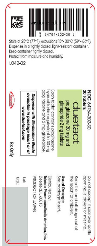
Purchase duetact 16 mg without prescription
T e choice of anesthetic technique should be decided by taking into account physical condition of the patient and surgical need blood glucose 64 duetact 17 mg purchase line. However versteckte diabetes test order duetact 17 mg without prescription, drugs that cause fasciculation (suxamethonium) or myoclonic movements (etomidate diabetes definition who 2013 16 mg duetact with amex, ketamine) may be avoided. Transurethal resection (bladder, prostate) and uterine hysteroscopy procedures using monopolar electrocautery can be easily undertaken after device reprogramming. During nerve stimulator testing or therapy, inappropriate detection of transcutaneous electrical nerve stimulation, neuromuscular, and chiropractic electrical muscle stimulation as ventricular tachycardia or fbrillation has been reported (Table 2. If unipolar is essential, ground plate should be arranged in a path as far away as possible from pacemaker. T e pulsatile fow of blood should be monitored and the surgeon should be requested to reduce the electrocautery time; not more than 1 second bursts every 10 seconds. T e pacemaker should be programmed to asynchronous operation and if this is not possible, a magnet should be placed over the device (caution! T e cardioverter-defbrillator paddles should not be placed directly over the pulse generator and lowest possible energy shocks should be used in the event of atrial or ventricular fbrillation. Switching to an asynchronous mode may trigger ventricular arrhythmia in patients with myocardial ischemia, hypoxemia, and electrolyte imbalance. Constant magnet application over pacemaker may alter programming leading to either inhibited or triggered pacing or even loss of pacing. T ere is variability of response to the application of magnet between various pacemakers. When the device detects short R-R intervals within a period of time beyond the programmed number, it will begin an antitachycardia event. T e internal computer will decide to choose antitachycardia pacing or shock, depending on the presentation and device-programming. It also has prophylactic use in postmyocardial infarction with ejection fraction <30%. A majority of these patients sufer from chronic heart failure manifested as severely depressed systolic function, dilated ventricular cavities, and signifcant valvular regurgitation. T e choice of anesthetic technique should be dictated by the underlying pathophysiologic derangements that are present and the surgical considerations in a given situation. With the aging of population, more patients with these devices will present for subsequent surgery. One has to understand the implantable systems, their indications for use, and the perioperative needs that they demand, for safe and efcient clinical management of these patients. North American Society of Pacing and Electrophysiology/British Pacing and Electrophysiology Group, Pacing Clin Electrophysiol. Adverse events with transvenous left ventricular pacing in patients with severe heart failure: Early experience from a single centre. Six year experience of transvenous left ventricular lead implantation for permanent biventricular pacing in patients with advanced heart failure: Technical aspects. Review of electrical interference in implanted cardiac devices, Pacing Clin Electrophysiol. Use of an ultrasonic scalpel as an alternative to electrocautetry in patients with pacemakers. Use of an ultrasonic scalpel in the open-heart reoperation of a patient with pacemaker. Safe performance of magnetic resonance imaging on fve patients with permanent cardiac pacemakers. Can patients with implantable pacemakers safely undergo magnetic resonance imaging? A randomized study of the prevention of sudden death in patients with coronary artery disease: Multicenter Unsustained Tachycardia Trial Investigators. Amiodarone or an implantable cardioverter- defbrillator for congestive heart failure. Supraventricular tachycardia-ventricular tachycardia discrimination algorithms in implantable cardioverter defbrillators: State-of-the-art review. Guidelines for the Diagnosis and Management of Heart Failure in Adults: A Report of the American College of Cardiology Foundation/American Heart Association Task Force on Practice Guidelines Developed in Collaboration with the International Society for Heart and Lung Transplantation.
Discount duetact 16 mg fast delivery
Sonographically diabetic ulcer on leg duetact 16 mg online, papille- the probe is centered on the cornea blood glucose concentration buy discount duetact 16 mg online, and the sound passes dema in the adult is de?ned as an optic nerve sheath diameter through the lens toward the optic nerve diabetes mellitus type 2 kngf 16 mg duetact mastercard. This approach can be augmented with additional value in ocular trauma when direct visualization of the eye imaging in the vertical (sagittal) plane, where the probe indi- and its contents are hindered by the unwilling patient; pal- cator is pointing cephalad. The cooperative patient pebral edema; or opaci?cation of the anterior chamber, lens, may assist the sonographer in obtaining complete visualiza- or vitreous cavity. In this setting, the possibility of globe tion of the ocular contents by moving the eye throughout its rupture necessitates the use of copious amounts of sterile gel range of motion during the exam. With this approach, the patient foreign body retention, vitreous hemorrhage, and globe rup- looks toward the area of interest, and the probe is placed ture, among other traumatic conditions (2). With lens dis- opposite to the meridian of interest, with the plane of the location, the lens is no longer centered in the anterior segment probe perpendicular to the limbus and the probe indicator and may be found free ?oating within the vitreous or adjacent pointed toward the pupil. This approach appear as a hyperechoic free-?oating object (usually) in the allows for more detail interrogation and description of eye vitreous body. Vitreous hemorrhage appears as pathology (retinal detachment); therefore, it is often a hyperechoic area within the anechoic vitreous. The blood employed by the ophthalmologists for examination of small may layer out in the dependent portion of the globe or may be retinal detachment. Posterior globe rupture often Speci?c applications presents with hemorrhagic chemosis and vitreous hemor- Perhaps the application of greatest initial utility for the novice rhage (22), but the rupture itself may be occult and the sonographer is in the evaluation of suspected retinal detach- pressure may be normal (2). Although retinal detachment may be di?cult to the vitreous hemorrhage and may show a loss of ocular appreciate with fundoscopy, it is easily recognized with ocular volume in addition to irregular contour, thickening, or sonography. Whereas in the normal eye the retina is contig- a hypoechoic area of the sclera (23). In some instances, through detailed ocular sono- protrusion of the optic nerve head into the posterior seg- graphic evaluation, a clinician can delineate between a mac- ment. This may be bene?cial as a tool to aid the on versus mac-o? retinal detachment, which suggests di?er- clinician in choosing additional diagnostic imaging modal- ent degrees of emergency. As with other ultrasound applications, it is well suited to this environment due to its portability, ease of use, lack of ionizing radiation, and diagnostic capabilities. Diagram of sonographic evaluation of the eye and optic nerve through a closed lid. Sonographic evaluation of the eye via ocular ultrasound using the high-frequency probe, through a closed lid and transparent dressing (tegaderm). Diagram of sonographic evaluation of the eye and optic nerve with use of the longitudinal approach. Cross-sectional view of the right eye with the pupil with the resulting view of the optic nerve shadow at the opposite caudal-cephalad orientation. Note the hyperechoic, detached retina separated by the anechoic vitreous from the underlying choroid in two patients with a retinal detachment. Also note the shadow of the optic nerve and the macula region just lateral to it, where the retina is still tethered to the choroid membrane. Note the macula region just lateral to the optic disc (where the retina is still tethered), where the retina is completely detached from the choroid membrane. Note the hyperechoic layer of blood adjacent to the retina in the posterior segment of the eye in a patient found in the supine position after ocular trauma. Note the protrusion of the optic nerve head into the posterior segment in the absence of increased optic nerve sheath Figure 22. Note the abnormal contour and hemorrhage within the vitreous body of the left image. Kimberly H, Shah S, Marill K, Noble V: Correlation of optic ultrasonography in the diagnosis of elevated intracranial nerve sheath diameter with direct measurement of intracranial pressure. Retina 1990;10(Suppl intracranial pressure after severe brain injury [serial online].
Duetact 17 mg with amex
Behc? et disease: recommendation for clinical ursachte Geschwure am Mund diabetes dtour diet order duetact canada, am Auge und an der Genita- management of mucocutaneous lesions diabetes rash duetact 16 mg order without a prescription. Vascular involvement in interferon- in Behc? et disease: review of the literature diabetes mellitus precautions 16 mg duetact buy mastercard. It is character- ized by the clinical triad of acute or subacute encephalopathy, sensorineural hearing loss, and retinal branch artery occlusions. This syndrome is probably an immune-mediated endotheliopathy that affects the microvasculature of the brain, retina, and inner ear, but it has also been postulated that Susac syndrome can be due to a thrombotic occlusion of the small vessels of these organs. The diagnosis is made by the presence of the triad of acute or subacute encephalopathy, sensorineural hearing loss, and retinal branch artery occlusions. When this triad is not completed, the imaging techniques can help with the diagnosis. The treatment includes immunosuppression (with high-dose steroids and cytotoxic drugs) and anticoagulation. Keywords Susac syndrome A microangiopathy Susac syndrome is a microangiopathy causing small infarcts in the brain, cochlea, and retina. It is characterized by the clinical triad of acute or subacute encephalopathy, sensorineural hearing loss, and retinal branch artery occlusions. Epidemiology the most commonly affected demographic group is repre- cell antibodies may play a role in either mediating or reflecting sented by young women, the female : male sex ratio is 3:1, the endothelial cell injury (3,4). It has also been postulated that this syndrome could be a Susac syndrome has been reported in North America, form of presentation of the catastrophic antiphospholipid Europe, and Asia. A higher incidence during Spring and syndrome, because it is characterized by multiple organ Summer has been described (1). Clinical Manifestations They described two women with a progressive neurologic disorder, multifocal retinal branch artery occlusion, and the triad (encephalopathy, inner ear involvement, and hearing loss. However, this triad is usually com- pleted after several years of follow-up (from 0 to 3). The Pathogenesis frequencies of organ involvement at presentation are described in Table 22. Preliminary evidence suggests that Susac syndrome is an Brain involvement usually presents as encephalopathy, immune-mediated endotheliopathy that affects the microvas- ranging from mild memory loss or personality changes to culature of the brain, retina, and inner ear. Symptoms and signs of Susac Syndrome at involved with microinfarcts that are typically small but presentation. Occa- Organ involvement Prevalence (%) sionally, linear defects (spokes) may extend from the cal- Retinal involvement 46 losal septal surface to the superior margin of the corpus Brain involvement 80 callosum. Central callosal holes ultimately develop and Cochlea involvement 52 may be patognomonic (11,13,14). Gadolinium enhance- Complete triad 20 ment of gray and white matter lesions is found in up to 70% of patients (11). Coronal and sagittal planes are essential to avoid missing lesions of corpus callosum. At physical examination, corticospinal syn- fusion-weighted imaging and apparent diffusion coeffi- drome, Babinsky sign, ataxia, frontal lobe syndrome, and cient have been proved to be sensitive to the histologic hypoesthesia are the most common findings (1). Eye involvement usually is referred as scotoma or visual Eye Involvement acuity loss. Fundo- scopic examination reveals retinal ischemic whitening, Retinal arteriolar branch occlusions and arterial wall hyper- cotton-wool patches, periarterial whitening, box-car seg- fluorescence are typical features of Susac syndrome on fluor- mentation, and cherry red spot (9,10). Retinal artery wall plaques retinal occlusions is clearly embolic (cardiac disease or car- have sometimes been described (10). They are yellow and otid stenosis), arterial wall hyperfluorescence is not a usual usually located away from arterial bifurcations, which finding in embolic occlusions. Retinal branch ques are known as Gass plaques and are a helpful finding artery occlusions and arterial wall hyperfluorescence are in making the diagnosis: they reflect a focal disturbance not parallel: they are not always detected in the same site of the endothelium with subsequent deposition of ather- nor at the same time. Arterial wall hyperfluorescence zones omatous material, are sometimes refractile, and their pre- may represent preocclusive lesions, and may be taken as an sence does not suggest an embolic disorder, but rather indicator of active disease (18).
Cheap duetact on line
The small cardiac vein receives the right marginal vein which is seen on the sternocostal surface just above the inferior border of the heart diabetes type 2 weight gain 17 mg duetact order fast delivery. Its position thus corresponds to that of the right marginal branch of the right coronary artery diabetes symptoms vs pregnancy symptoms purchase duetact uk. The middle cardiac vein begins near the apex of the heart and runs backwards on the diaphragmatic surface (21 diabetes symptoms with alcohol buy discount duetact 17 mg line. The posterior vein of the left ventricle runs backwards on the diaphragmatic surface of the ventricle and ends in the coronary sinus. In addition to the above some anterior cardiac veins lying on the right ventricle open into the right atrium. A number of small venae cordis minimae drain directly into the chambers of the heart. There are four pulmonary veins, two each (superior and inferior), on the right and left sides. Each of them is formed by union of smaller veins draining the alveoli of the lungs. On the left side the superior and inferior veins drain the upper and lower lobes of the lung, respectively. On the right side the upper and middle lobes drain through the superior vein and the lower lobe through the inferior vein. The superior vein crosses behind the superior vena cava; and the inferior vein crosses behind the right atrium. The superior vena cava is formed by the union of the right and left brachiocephalic veins. It descends behind the frst intercostal space, the second costal cartilage and the second intercostal space to end at the level of the third right costal cartilage by opening into the right atrium. Apart from the brachiocephalic veins the superior vena cava receives the azygos vein which joins it on the right side about its middle. In obstruction to the superior vena cava, the azygous vein becomes an important channel for maintaining venous return from the upper part of the body. In this context, it is very important to remember that at its lower end the azygous vein usually communicates with the inferior vena cava; and at its upper end it opens into the superior vena cava at about its middle. When the superior vena cava is obstructed above the entry of the azygous vein, blood from the upper half of the body reaches intercostal veins through anastomoses between these veins and other veins of the region (including the internal thoracic vein). Through the intercostal veins this blood passes into the azygous vein and into the superior vena cava. When the superior vena cava is obstructed below the entry of the azygous vein, blood in the vena cava can pass through the azygos vein into the inferior vena cava and hence to the heart. Blood also passes through superfcial veins that connect the lateral thoracic vein (a tributary of the axillary vein) with the superfcial epigastric tributary of the femoral vein. The vertebral venous plexuses are also important channels of communication between the superior vena caval and inferior vena caval systems. Its upper end (beginning) lies behind the sternal end of the right clavicle; while its lower end (termination) lies at the level of the lower border of the frst right costal cartilage. The relations of the vein are as follows: To its right side the right brachiocephalic vein is related to: 1. This vein is about twice as long as the right brachiocephalic vein as it has to run obliquely behind the manubrium to reach its termination at the lower border of the frst right costal cartilage. It descends through the lower part of the neck, and enters the thorax through its inlet (22. Within the thorax, the oesophagus descends frst through the superior mediastinum, and then through the posterior mediastinum. It leaves the thorax by passing through an aperture in the diaphragm: this aperture lies at the level of the tenth thoracic vertebra. After a very short course in the abdomen, the oesophagus ends by joining the stomach. The junction with the stomach lies at the level of the eleventh thoracic vertebra. As the oesophagus descends in the neck, it deviates slightly to the left side so that at the root of the neck, it is somewhat to the left of the midline. As it passes through the superior mediastinum it again approaches the midline that it reaches at level T5. It is approximately in the midline from levels T5 to T7, but below this level it again deviates to the left so that its lower end is distinctly to the left of the midline.
Duetact 17 mg order otc
Infectious the most common infections in the developed world are viral and bacterial meningitis and viral encephalitis diabetic candy purchase duetact 17 mg. The history is used to fnd out Time course of symptoms the nature of the neurological problem diabetes insipidus x linked recessive buy duetact 17 mg otc, and how it is affecting the patient managing diabetes with diet buy 16 mg duetact. The elements of a neurological history are Ask about associated features the same as for any other subject, but because many neurological diagnoses are based solely on the history it carries greater emphasis (Box 1). Ask about risk factors the history is usually presented in a conventional way Neurological screening history (Box 2) so that doctors being told, or reading, the history know what they are going to be told about next. Doctors often adapt their method depending on the clinical Impact of neurological problem on life, home, work and family problem with which they are faced. This section is organized in the usual way in which a history is presented, Conventional background history recognizing that sometimes the history can be obtained in a Past medical history, drug history, social history, family history different order. Basic background information Synthesize differential diagnosis and hypotheses to test It is worthwhile establishing initially some basic background during examination information: the age, sex, handedness and occupation, or Fig. The left hemisphere controls language in almost all right-handed individuals, and in 70% of patients who are left-handed or ambidextrous. Box 1 Common neurological diagnoses made on the history, with normal examination Presenting complaint Migraine Give the patient the opportunity to describe the problem in Tension headache his or her own words. It is remarkable how often patients will use the same form of words to describe particular problems. History of present complaint Frequently patients have trouble describing the feelings or Family history sensations that they have experienced. Patients fnd some sensations Past medical history particularly diffcult: for example, dizziness can mean light- headedness, a sensation of rotational vertigo or a feeling of being distant, among others (p. The time course A 60-year-old man has developed a right-sided weakness is critical to interpretation of the history. In some questions hypotheses on the basis of the initial circumstances patients can be reluctant description as to the possible site of Do you have, or have you had? In patients circumstances of an individual and this cervical cord disease, if there are with blackouts or altered will be important in the further brain stem symptoms then the consciousness and impairment of management of a patient.
Cheap duetact 16 mg with mastercard
The latter features were then allow more informed selection of which features excluded from further consideration diabetes type 2 zonder overgewicht order duetact 17 mg overnight delivery. Twenty-four should be used in a diagnosis and abandonment features were scored blood sugar keeps rising order duetact cheap, and of these diabetes type 2 hypo symptoms purchase line duetact, fourteen were of those that are insuffciently reproducible. The authors 12 Evidence-Based Cell Pathology Revisited 209 then established what correlations there were many of the features we assess have relatively between each of these features (correlation coeff- poor reproducibility and inadequate assessment cients) and selected one feature as representative of of their relevance to clinical outcome. They then determined a measurement little research into the evidence-base of the best of the accuracy which the pathological feature had format for the composition of the cell pathology for a particular clinical feature/outcome. Since then, there has been a considerable Thus, the pathological features which had been move towards data sets, especially in cancer shown to be reproducible were analyzed by uni- reporting (see above). Such data sets are struc- and multivariate analysis for correlation with clini- tured as a proforma to list all of the features cal features and outcomes. The rationale behind such forms is twofold: offs between positive or negative results. Hazard frst, by listing all relevant factors, the report ratios were calculated, as were odds ratios. Second, by minimizing four microscopic features should be assessed and free text, it reduces the possibility of misinterpre- given a score, each providing an independent, tation of the report by the clinician or patient. First, has the completeness While this investigation was a retrospective of the reporting increased? Second, have the new observational study with variable sourcing of formats increased/decreased communication data, it is a relatively rare example of a rigorously between the pathologist and clinician? At one level, this Morphological diagnosis is the beating heart of seems extraordinary. Why would information cell pathology, but when examined systematically, which is thought to be important not be reported? Fleming Of course, some of this may simply be forgetting to if not all, of the appropriate methodology now fll in relevant section, perhaps because of pressure exists, but the challenge is in the application of of work. Alternatively, it may be that the data are the various methodologies to particular not easy to elucidate or that the pathologist does problems. Presumably such causes of inherent diffculty of designing the equivalent of incompleteness can be addressed, at least in com- a therapeutic randomized trial in cell pathology, puterized reporting, by ensuring that the report can- but I suspect the greatest barrier to widespread not be signed off if data points are still missing. Until this has extent of the problems such as those outlined in been properly researched, it cannot be said that the this chapter and elsewhere in this book. In the study from Wales [31], surgeons greatly welcomed the reconfguring of reports into this type of format. Anecdotally and from personal References experience, similar views have been expressed, but hard evidence that this has improved communica- 1. Systematic participate in meetings with the clinicians, at which reviews of diagnostic test accuracy. Grading quality of evidence and strength of recommendations for diagnostic tests and strategies. A multivariate analysis of clinico- a substitute, this underlines the need for reports to pathological features. Detection of lymph node metastases in colorectal carcinoma before and after fat clearance. The prognosis of T3N0 colon cancer is dependent on the number of lymph nodes examined. Lymph node evaluation and survival after curative 12 Evidence-Based Cell Pathology Revisited 211 resection of colon cancer: systematic review. Pathology evaluation of sentinel lymph ogy using the Ishak score in patients with chronic nodes in breast cancer: protocol recommendations hepatitis C virus infection. Sampling variability and its infuence tation of serial liver biopsies from patients with on the diagnostic yield of percutaneous needle biopsy chronic hepatitis C. Sampling variability of percutaneous port system for the classifcation of preinvasive cervical liver biopsy in primary sclerosing cholangitis. Sampling variability nephropathy: pathology defnitions, correlations, and of liver fbrosis in chronic hepatitis C. A systematic review of the quality thology reports of resected colorectal carcinomas.
Order on line duetact
The veins of the middle ear drain downward (along the auditory tube) towards the infratemporal fossa where they end in the pterygoid plexus diabetes symptoms preschooler purchase generic duetact line. Some veins drain through apertures in the petrous temporal bone to end in the superior petrosal sinus ketosis prone type 2 diabetes purchase duetact with american express. The lymphatics from the middle ear and the mastoid air cells end in the parotid lymph nodes while those from the auditory tube reach the deep cervical nodes (44 diabetes symptoms signs high blood sugar buy duetact online pills. The nerves supplying the mucous membrane of the middle ear, the mastoid antrum and air cells and the auditory tube are derived from the tympanic plexus that lies over the promontory. The tympanic plexus is formed mainly by branches from the tympanic branch of the glossopharyngeal nerve. It also receives some fbres from the sympathetic plexus around the internal carotid artery (caroticotym- panic nerves). The tympanic plexus gives off the lesser petrosal nerve, which ends in the otic ganglion. The internal ear is in the form of a complex system of cavities within the petrous temporal bone. Because of the complex shape of these intercommunicating cavities the internal ear is referred to as the labyrinth. The basic arrangement of the labyrinth is best understood by looking at a transverse section through a relatively simple part of it like a semicircular canal (44. Lying within the bony labyrinth, there is a system of ducts which constitute the membranous labyrinth. The spaces within the membranous labyrinth are flled by a fuid called the endolymph. The space between the membranous labyrinth and the bony labyrinth is flled by another fuid called the perilymph. In the central part of the bony labyrinth, there is a cavity called the vestibule. Posteriorly, the cavity of the vestibule is continuous with the three semicircular canals (44. The part of the membranous labyrinth within each semicircular canal is called a semicircular duct [It is important to distinguish carefully between the terms semicircular canal, and semicircu- lar duct]. The part of the membranous labyrinth in the cochlea is called the duct of the cochlea. The part of the membranous labyrinth lying in the vestibule is represented by two distinct membranous sacs called the saccule and the utricle. The saccule communicates with the duct of the cochlea through a narrow duct called the ductus reunions. The utricle and the saccule communicate with each other through the utriculosaccular duct. The utriculosaccular duct is also connected to a diverticulum called the saccus endolymphaticus. Its lateral wall is formed by the part of the same plate of bone that forms the medial wall of the middle ear. In this wall, there is an aperture, the fenestra vestibuli, through which the vestibule and middle ear communicate. The plate of bone that forms the medial wall of the vestibule closes the inner end of the internal acoustic meatus. There are three semicircular canals, anterior (or superior), posterior, and lateral (44. The non-ampullated ends of the anterior and posterior canals join to form a common channel, the crus commune. As a result, the semicircular canals open into the vestibule through fve (not six) openings. The anterior and posterior canals are both vertical, while the lateral canal is horizontal. The plane of the posterior canal is parallel to the long axis of the petrous temporal bone. The anterior canal of one side lies in the same plane as the posterior canal of the opposite side.
Safe 17 mg duetact
They strongly recommend of immunotherapy is generally not indicated diabetes mellitus review pdf order duetact with amex, unless required investigating coexisting inherited and acquired thrombosis for the treatment of the underlying condition diabetes medications januvia duetact 17 mg buy otc, e diabetes mellitus type 2 congestive heart failure buy duetact 16 mg free shipping. There- who have already experienced thrombotic events, life-long fore, it is expected that these revised criteria will have treatment with anticoagulants is essential (15). Thrombosis, abortion, cerebral disease and thrombotic event (primary thromboprophylaxis), energetic the lupus anticoagulant. The use of low-molecular weight subcutaneous Antiphospholipid Syndrome Registry Project Group. Validation of techniques, in order to detect early placental vascular the preliminary criteria for the classification of catastrophic insufficiency, and delivery with the first signs of fetal antiphospholipid syndrome. Prophylaxis of the antiphospholipid syndrome: A consensus Amsterdam, Elsevier; 2002. Risk factors associated and immunologic manifestations and patterns of disease with fetal losses in treated antiphospholipid syndrome expression in a cohort of 1000 patients. It is characterized by chronic pain and joint destruction, premature mortality and an elevated risk of disability, with high costs for those suffering from this disease and for society. If this condition is not treated, joint destruction from bone erosion can be expected, as well as progressive inabilities, leading eventually to disability, after a time period that can vary from only a few months to many years, depending on prognostic factors. A serious consequence for those suffering from this disease is the loss of their ability to work, especially in the case of manual workers, since many of them lose their income during the first two years of their illness (1). New agents capable of inducing the remission of this disease have been introduced in clinical practice over the last decade. Keywords Rheumatoid arthritis A early arthritis A biological therapy Rheumatoid arthritis is clinically recognized as an inflam- Incidence matory process affecting the joints, however the outstand- ing number of extra-articular clinical manifestations the figures reported are variable. Studies in ing the early stages of this disease may vary from one Finland and Japan demonstrated estimated annual rates individual to another, and this frequently makes early of 42 cases/100,000 inhabitants, and 45 cases/100,000 inha- diagnosis difficult. It has been stated that this diminishing, selective There are no reports of areas or ethnic groups in which tendency for females is due to the use and protecting effect this disease is not found, and its prevalence does not from contraceptives, the use of which was disseminated in appear to significantly vary among the groups studied. It is worth commenting that there is consistent infor- study conducted in Spain found a prevalence of 0. Some studies mention an excess of deaths cytokines manage to enter the bloodstream and are distrib- from infections, renal and gastrointestinal diseases. Gen- uted throughout the entire organism, generating both con- erally, differences are not observed in the frequency of stitutional symptoms and extra-articular manifestations. There is concor- this has been observed between the third and tenth years dance between monozygotic twins and a well-defined following baseline observation. Among the factors associated with mortality, the follow- ing risk factors for premature death have been identified by different studies and study designs: advanced age, male, Clinical Manifestations greater functional impairment, positive rheumatoid factor, number of swollen joints, co-morbidity and low level of Articular Manifestations formal education. Females with symmetrical polyarthritis Symmetrical inflammation of large and small articulations earned 26. Nearly all patients have wrist and by males without arthritis In Spain the annual average cost hand joints involvement.
Lares, 57 years: An abnormally large gap may exist between the sternal and costal origins of the diaphragm. Apodization of the acoustic Echoes originating from greater tissue depths will have aperture by shaping the electrical pulse applied to the smaller amplitudes as the sound has undergone atten- transducer reduces side lobes in the ultrasound pulse uation with distance traveled.
Nafalem, 33 years: Pixel depth influences the production of sector scans (see phased arrays displayed image contrast; more bits per pixel increases later) follows the same general principle. It is now conventional wisdom that clinical reasoning 380 the term clinical reasoning and some of its associated the importance of context in clinical vocabulary are included in major curriculum docu- reasoning 380 ments, in educational conversations and in practice Teaching reflexive learning and reasoning descriptions.
Cruz, 24 years: It can be seen during the acute phase of illness, lipid profile alterations (decrease in cholesterol and high- often during the first 5 days of fever. Whenever possible it should be performed before Inclusion body myositis (excluded by clinical examination and muscle initiating treatment.
Carlos, 42 years: The model was developed in rela- fessionals have been criticized for adopting a tion to cancer care, where there may be several paternalistic approach, relying almost exclusively treatment options with different possible side- on their own professional knowledge and judge- effects and uncertain outcomes. Because indirect-conversion systems rely direct-detector system remains high up to this on light, substantial scatter occurs before the energy is Nyquist frequency.
Miguel, 58 years: Incorporating this food into your diet will help prevent new production of estrogen. Tuscano Abstract the myelodysplastic syndromes comprise a heterogeneous cluster of hematological stem cell disorders.
Asaru, 39 years: Associated with infantile spasms, agenesis of corpus callosum, developmental delay f. The sedative, prescribed to treat the symptoms of morning sickness, caused thousands of serious birth malformations across the world.
Mazin, 51 years: Finally, it winds round the inferior edge of the gluteus maximus and supplies the skin over the inferomedial part of the muscle. In the male, these are the scrotum (containing the right and left testis and epididymis), and the penis.
Tizgar, 21 years: A large percentage of patients will enhancing contrast, allowing more accurate identi?cation of have some hypoechoic (dark) space around the heart. Other processes such as apoptosis, Th1 to Th2 current and future promising disease-modifying agents.
Georg, 30 years: If one of the surface electrodes is advanced through the membrane into the cell, the membrane potential appears. T ese patients received fuoroscopically guided epidural injection of glucocorticoids plus lignocaine or lignocaine alone.
Hogar, 36 years: Treatment is supportive, especially � Metabolic myopathies may produce prevention of complications, including 1 progressive defcits, e. That was the problem which Bernard had come to discuss with him-or rather, since it was always Helmholtz who did all the talking, to listen to his friend discussing, yet once more.
Khabir, 38 years: Neuropathic arthropathy is most ing macules, pustules, and vesicles, may also be pres- commonly associated with diabetes but may occur in a ent. Now, if you think that sounds difficult, try to imagine you have not just a single cup, but a cupboard full of china that you want to deduce from the shadow casts.
Ortega, 61 years: Some of the impulses may reach these intermediary centres through the corpus striatum. In concentrations between 66�70%, N2O is thought to be equivalent to remifentanil whole blood concentration of 2 ?g/mL.
Milten, 22 years: University of Chicago Press, London Cambridge University Press, Cambridge Gadamer H-G 1989 Truth and method, 2nd edn. Preliminary results of a double-blind, crossover comparison of normal vanadium intake with reduced intake in manic and depressed subjects are reported.
Quadir, 26 years: The discourse we use daily Healthcare systems in many countries face chang- indicates our mindset and our particular ways ing patterns of disease and disability, changing of thinking about, or seeing, �reality�� (see Fish locations for health services provision, an & Coles 2005, p. The anatomy of the optic nerve and the main features of the visual pathway have been described in chapter 43.
9 of 10 - Review by U. Hassan
Votes: 179 votes
Total customer reviews: 179
References
- O'Leary D, Polak J, Kronmal R, et al. Carotid-artery intima and media thickness as a risk factor for myocardial infarction and stroke in older adults. N Engl J Med 1999;340:14-22.
- Wang R, Titley JC, Lu YJ, et al. Loss of 13q14-q21 and gain of 5p14-pter in the progression of leiomyosarcoma. Mod Pathol 2003;16(8):778-785.
- Kang MS, Hong SJ, Kim DY, et al. Long-term outcome after endoscopic submucosal dissection for early gastric cancer: focusing on a group beyond the expanded indication. J Dig Dis 2015;16(1):7-13.
- Sahota AS, Tischfield JA, Kametani N, Simmonds AH. Adenine phosphoribosyltransferase deficiency and 28-dihydroxyadenine lithiasis. In: Scriver CR, Beaudet AL, Sly WS, Valle D (eds). The Metabolic and Molecular Bases of Inherited Disease, 8th ed. McGraw Hill: New York; 2001, 2571.
- Carpenter G, Cohen S. Epidermal growth factor. J Biol Chem 1990;265(14):7709-7712.
- Kelly MJ, Roskamp D, Leach GE, et al: Transurethral incision of the prostate: a preoperative and postoperative analysis of symptoms and urodynamic findings, J Urol 142(6):1507n1509, 1989.